Functional Connectivity Mapping in Preoperative Neurosurgical Planning: Advances to Improve Surgical Outcomes
Preoperative planning in neurosurgery can sometimes be exhaustive. Functional connectivity mapping also secures an important place there. The technique will help the doctors to locate those areas of the brain well in advance of surgery that are truly vital. It is the thought of how various areas of the brain communicate with each other that may further assist surgeons in working around or entirely avoiding critical areas and thereby minimize risks during the course of a procedure, improving recovery afterward.
This is done by a mapping process with the use of high-level imaging that outlines the connections in the brain. Analysis of these pathways provides the surgeons with the development of particular strategies for their patients, which enhances safety and efficacy during the operations. The knowledge gleaned from mapping guides decisions that benefit patient care.
These tools must be espoused by surgeons and their medical teams so that surgical planning could be enhanced. The insights afforded by functional connectivity mapping are truly changing the face of neurosurgery, with patient safety and effectiveness of surgery right at the top of the list.
Key Takeaways
- Functional connectivity mapping helps identify the essential regions of the brain prior to surgery.
- State-of-the-art imaging techniques visualize the connections of the brain for an optimized surgical strategy.
- Tailored approaches based on mapping lead to improved patient outcomes and safety.
Principles of Functional Connectivity
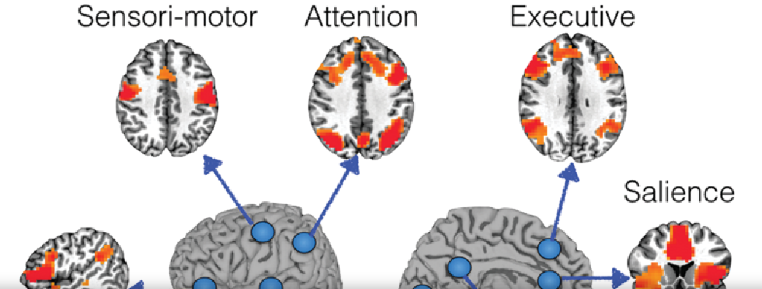
It is the pattern of interaction between different regions of the brain. The idea of such connections is vital in many fields, but most importantly in preoperative neurosurgical planning. Key theories and technologies help in mapping such connections effectively.
Neural Network Theories
Neural network theories describe how different areas of the brain communicate with one another. The theories argue that functional connections form networks of interacting neurons. In other words, it is argued to mean that most of the activities in the brain are done in collaboration between multiple regions; thus, an activity in one area might influence another.
For example, the DMN is a resting state network that involves thought. Research in this field allows for an advanced understanding of the functions of these networks in health and disease. It’s by the knowledge of such networks that surgeons are able to identify areas to avoid when performing surgery.
Functional MRI and Its Role
Functional MRI represents one of the critical tools for brain activation studies. It relies on the principle that changes in blood flow associated with neural activity should be detectable: the more active an area of the brain, the more oxygen it requires, which is precisely what fMRI detects.
This technique helps in the identification of functional connectivity by showing cooperating areas of activation in specific tasks. Surgical teams use data from fMRI to help plan safe approaches to brain surgeries. It reveals key connections which must not be disturbed by any surgical intervention and often improves outcomes for patients.
Graph Theory in Connectivity Analysis
Graph theory describes brain networks using a mathematical framework. In the context of the brain, nodes represent brain regions and edges depict the connection between these. Such methodology will go a long way in assisting researchers in exploring how information flows within the brain.
Graph theory metrics may give further insight into the strength of connectivity and efficiency of such networks. For instance, connectivity maps may highlight the presence of hubs or key interconnecting points in such networks. This can be very important to neurosurgeons, who would want to avoid damaging such critical regions of the brain during a surgical procedure.
Preoperative Functional Mapping
Preoperative functional mapping enables neurosurgeons to understand the activity of the brain prior to the operation. It pinpoints the imperative areas responsible for controlling functions such as movement, speech, and memory. Mapping using different techniques thus guides surgical procedures by minimizing risks.
Techniques for Eliciting Functional Areas
Techniques that have been employed to map functional areas of the brain vary. The most common technique used is Functional Magnetic Resonance Imaging, fMRI; it points to regions of the brain which are active when subjects perform specific tasks.
Another successful approach is Electrocorticography, or ECoG. This is a method in which electrodes are placed directly on the surface of the brain to measure electrical activity.
Transcranial Magnetic Stimulation is also able to stimulate brain regions non-invasively. Each one of these techniques has some advantages and disadvantages depending on the case.
These methods ensure that surgeons are able to conceptualize and identify essential functions which will guide their approach to surgery.
Integration into Surgical Planning
It’s crucial to integrate functional mapping into surgical planning. Neurosurgeons study the maps to find out areas responsible for vital activities. This helps in avoiding such areas during surgery.
Detailed maps influence the surgical approach. They also affect the estimated recovery process: if surgeons know what the crucial areas are, they can employ techniques that disturb the functions the least.
Coordination for success by teams requires the cooperation of neurologists, radiologists, and surgeons. Effective communication allows assurance that the surgical plan reflects the functional architecture of the brain.
Case Studies: Successes and Challenges
Real-life case studies bring into the limelight the importance of functional mapping. There is this patient who had undergone surgery for a tumor near his speech center. Precise location by preoperative mapping allowed the surgeon to successfully remove the tumor, which had least effects on speech.
Another example is the discovery of unexpected functions in areas that were assumed to be non-critical, which then required intra-operative adjustments in surgical plans, hence requiring some flexibility.
These examples stress the fact that, while functional mapping very much helps in planning, it simultaneously brings about challenges. Each case is different, requiring careful evaluation for optimal outcomes.
Also Read :