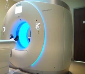
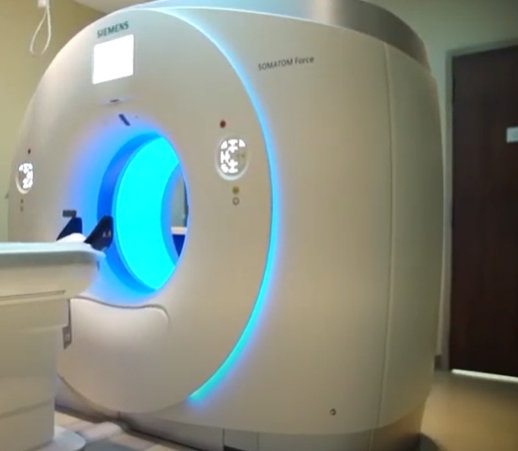
Intraoperative MRI: Revolutionizing Precision in Neurosurgery
Intraoperative MRI is one of many changes that are taking place in neurosurgery. With this new technology, surgeons take live pictures of the brain while performing surgery to help them better find and remove the tumor. Intraoperative MRI is very effective in improving surgical precision and thus improving outcomes for patients.
The surgeon can view directly the result of his work while still in the operating theatre. This capability does not only aid in making decisions that are uninfluenced at that instant, therefore reducing chances of complications and additional procedures later on. Many patients are able to have shorter recovery times and improved overall results.
In turn, according to the rise of this technology, neurosurgery stands a better chance of facing a future where operations will be a lot safer and more effective. Surgeons are, thus, readier to undertake more complex cases and push the envelope of what has been thought possible in brain surgery.
Key Takeaways
- Intraoperative MRI provides real-time imaging during the course of brain surgery.
- The use of this technology enhances the exactitude of tumor removal.
- Patients benefit with improved outcomes and quicker recoveries.
Advancements in Neurosurgical Procedures
Neurosurgery has dramatically changed with new imaging technologies. With the advancement, neurosurgeons may perform operations with a great degree of accuracy and safety as was never possible before.
History of Neuroimaging
Neuroimaging has evolved from the days when just simple CT and static MRI were used in practice. Though CT and MRI had their value, they did not offer updated information at the time of surgery. Early neuroimaging often led to some complication as information may be incomplete.
A very transformative period came with the introduction of intraoperative imaging. Surgeons now began to adapt MRI during operations, hence confirming tumor removal and integrity of the brain on the spot; no doubt this reduced second surgeries.
With these technologies, neurosurgeons rapidly saw a big improvement in patient outcomes. The live visualization of tumors and structures of the brain significantly changed surgical approaches.
Technological Advancement of MRI
Advancing technology in MRI has allowed for higher-quality imaging to be produced in shorter scan times. Newer MRI machines have more powerful magnets and advanced software, thus enabling them to provide sharper images of the brain.
Intraoperative MRI systems are now smaller and can be better accommodated into operating rooms. Technologies such as real-time imaging can enable surgeons to quickly make decisions on procedures. This capability minimizes risks and maximizes precision.
Moreover, better software enables greater delineation of cerebral tissues, tracts of nerves, and lesions. Better differentiation of normal and pathological tissues intraoperatively is possible. On the whole, these improvements lead to more successful interventions with a reduced complication rate.
Operative Advantages of Intraoperative MRI
Intraoperative MRI has a number of operative advantages in neurosurgical procedures. Improved accuracy, correction of brain shifts, and identification of vital structures are salient features of intraoperative MRI that contribute to patient safety and improved surgical outcomes.
Increased Accuracy in Tumor Resection
Intraoperative MRI is a critical modality that has greatly increased the accuracy of tumor resections. Surgeons can visualize the brain in question in real time during the operation. The immediate availability of such information would enable them to evaluate exactly how much tumor is left behind.
With this technology at their disposal, surgeons will be able to make speedy adjustments in approach. Indeed, studies show that complete tumor removal rates are higher with the use of intraoperative MRI. This reduces the chance of recurrence and can lead to improved long-term survival rates among patients.
Compensating for Brain Shift in Real Time
The brain shifts during a surgical operation, especially when some parts of the tumor are being taken out or if there is an alteration of blood flow. Such issues can be addressed through intraoperative MRI. Updated images provided by the technology reflect such changes.
In turn, these insights can be used by surgeons to refine the intraoperative surgical strategy in real time. In other words, by providing such information, these techniques will allow surgeons to more precisely target areas of tumor and avoid healthy tissue. This greatly enhances safety in the procedure.
Identification of Critical Structure
Intraoperative MRI helps to identify some critical structures of the brain. Some of these critical structures include nerves and blood vessels essential for living. By being able to visualize these areas, surgeons then can make appropriate decisions during operations.
This technology provides a safer way of doing multiple approaches to complicated brain tumors. This minimizes the risks of important tissue damage. The identification of these critical areas during surgery means better outcomes for patients since vital functions would have been preserved.

Also Read :
- Advanced MRI Techniques in Neurosurgical Planning
- The Role of Functional MRI in Neurosurgery
- The Intersection of Biomedical Engineering and Healthcare Innovation
- Tech Innovations in Healthcare: Improving Patient Care with AI
- Biomedical Engineering Projects: Innovative Solutions for Healthcare
