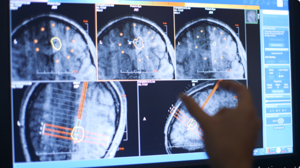
MRI Applications in Minimally Invasive Neurosurgery: Progress and Advantages MRI is beginning to revolutionize neurosurgery. It offers highly detailed pictures of the brain and spinal cord, thus allowing surgeons to attain a high degree of accuracy both in planning and executing surgeries. This technology enables safer and more effective minimally invasive procedures, hence reducing patients’ recovery times.
With the development of imaging, MRI already plays an important role in many surgical applications, enabling surgeons, for instance, to target areas of concern in real time during operations while sparing healthy tissue. The introduction of MRI into neurosurgical practice makes neurosurgery much less traumatic for the patient and much more efficient.
The continued demand for good treatment means that medical practitioners and patients alike have the answer they deserve over how MRI augments neurosurgery. This could further improve outcomes for patients and an appreciation of betterment in medical technologies.
Key Takeaways
- MRI provides neurosurgery with high-resolution pictures of the brain.
- It enables real-time guidance for minimally invasive procedures.
- MRI improves postoperative outcomes of the patient.
Basics of MRI in Neurosurgery
Neurosurgery depends on the integral use of Magnetic Resonance Imaging or MRI. This modality provides highly detailed images that assist in the diagnosis of pathology and planning of appropriate surgical intervention. Topics to be discussed will include the physical principles of MRI, current technological advances in the field, and how these features affect preoperative planning.
Principles of Magnetic Resonance Imaging
MRI utilizes powerful magnets and radio waves to produce detailed images of the brain and nervous system. When a patient lies inside the MRI machine, the latter produces a strong magnetic field. This field aligns hydrogen atoms in the body.
Then, the radiofrequency pulses disturb such alignment. Once such pulses are turned off, hydrogen atoms return to their previous positions while releasing energy at the same time. A computer captures the energy and converts it into images. These are high-resolution scans that outline tissues, tumors, and all abnormalities in the brain.
Advances in MRI Technology
Advances in MRI technology have enhanced its efficacy in neurosurgery several-fold. For instance, the new techniques being developed include functional MRI, which provides a view of the activities of the brain in real time. It develops an understanding of the patterns that different parts of the brain use and is also crucial during surgery.
With the development of many other techniques, like DTI, better visualization of white matter tracts is possible, thereby enabling the surgeons to avoid those areas. Enhancement in clarity and speed of images also reduces the time a patient has to spend inside the machine making all that more comfortable.
Preoperative Planning
Successful neurosurgery depends on preoperative planning. MRI views give surgeons the explicit images they need, helping them to locate the size and site of a tumor or another abnormality. Well-planned surgery is thus made possible by the acquisition of such information.
These surgeons can, with the aid of MRI scans, plan out the surgical procedure in their minds by mapping the best approach. They can also share such images with the surgical team for them also to understand the plan of action. When surgeons are well prepared, the possibilities of a positive outcome increase manifold.
Application of MRI in Surgical Procedures
In this regard, MRI is a significant tool in minimally invasive neurosurgery. It improves precision, safety, and results for a wide range of procedures. The key applications are: tumor resection, cerebrovascular surgeries, spinal surgeries, functional procedures, and postoperative analysis.
Brain Tumor Resection
MRI facilitates resection of brain tumors. The procedure generates 3-D images with information on tumor size and location. Surgeons base their approach to minimize damage to good tissue.
Intraoperative MRI can also give real-time imaging during the time of surgery. It allows for an assurance of complete tumor removal while preserving functional eloquence, such as speech and movement. This intraoperative guidance allows for the patient to not have to do additional follow-up surgeries afterward.
Cerebrovascular Surgery
In cerebrovascular procedures, MRI enables the visualization of blood vessels and points to blockages or even aneurysms. High-resolution pictures given by MRI assist surgeons in assessing the health of the vascular system.
In that respect, MRI angiography is particularly effective. It depicts the vessel structures and blood circulation in great detail, without the use of invasive procedures. This also serves very well for the exact planning of interventions, such as bypass or stent implantations.
Spinal Surgery
In spinal surgery, MRI helps in the diagnosis of herniated discs or tumors in the spine. These detailed images show surgeons the best approach route.
In this procedure, MRI may monitor the instrument position and confirm the access to the appropriate area of the spine. This minimizes complications and improves patient outcomes. Scans obtained post-surgery also contribute to the assessment of healing and complications.
Functional Neurosurgery
Functional neurosurgery addresses disorders like epilepsy and Parkinson’s disease. In each case, MRI provides a significant contribution to mapping brain regions responsible for functions such as movement and sensation.
By knowing the activity of the brain, surgeons are able to better target their treatments. MRI also offers the ability to monitor over time how treatments affect brain function to ensure optimal outcomes.
Postoperative Evaluation
Postoperative MRI is an important modality in the assessment of surgical outcome. It allows the investigation of residual tumor tissue and complications, which offers important information for subsequent management.
This will help surgeons to pick up problems such as infection or bleeding rather quickly and intervene in time. Regular MRI can also reassess the stages of recovery and further guide treatment if necessary.
Also Read :