
Quantitative MRI in Neurosurgical Risk Assessment: Improvement of Surgical Decision and Patient Outcome
Quantitative MRI is changing the way neurosurgeons assess risk before operating. It is an advanced imaging modality that avails critical information which may outline complications and, as such, helps in improving the outcomes. By measurement of the properties of the brain tissue, surgeons are informed and hence make decisions based on specific patient needs.
Applications of quantitative MRI in a clinical setup will serve to further understand the brain condition. This not only helps in diagnosing the condition but also leads to proper surgical planning for the neurosurgeon. A neurosurgeon applying this technology is better positioned to foresee risks and totally complex cases.
With the advent of quantitative MRI at virtually every doorstep, its application for neurosurgical risk assessment will increase. To understand principles and applications is the clue that enhances surgical precision, ensuring the safety of the patients.
Key Takeaways
- Quantitative MRI enhances information provided on brain tissue.
- Quantitative MRI neuroimaging modality improves neurosurgical procedures and patient outcome
- The increasing application of quantitative MRI becomes an indispensable and vital part of developing neurosurgical procedures.
Principle of Quantitative MRI
Quantitative MRI is essential for understanding the anatomy and functions of the brain. It can provide quantitative parameters that may be used to quantify risk factors prior to neurosurgery. This chapter introduces the technology and principles in imaging and biomarkers that are currently employed in quantitative MRI.
Neurosurgery and MRI Technology
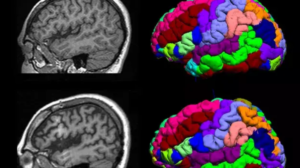
MRI technology uses very strong magnets combined with radio waves to produce highly detailed pictures of the brain. High-resolution imaging is usually necessary for neurosurgery to localize tumors, lesions, or other abnormalities.
The types of MRI most commonly used include:
- T1-weighted: Very useful when reviewing anatomy.
- T2-weighted: Good for lesion detection
- Diffusion-weighted imaging (DWI): Shows tissue integrity.
These imaging techniques play a critical role in offering information about the location and characteristics of brain issues. Advanced MRI machines would then use faster scans with improved image quality to enhance surgical planning and outcomes.
Principles of Quantitative Imaging
Quantitative imaging involves measurement of specific properties of tissues. In contrast to the standard MRI, which does not offer numerical data that quantify abnormalities in the brain, some of the key principles include:
Signal Intensity: This measures properties of tissues based on strength or amplitude of the MRI signal.
Relaxation Times: T1 and T2 times are the ways of measuring how fast the protons go back to equilibrium once disturbed.
These measurements help differentiate between healthy areas and diseased tissues. Protocols being standardized make it easier to compare results of one scan from another.
Biomarkers in MRI Assessment
Biomarkers represent those measurable indicators obtained from MRI scans. Biomarkers yield valuable insights into the state of brain health. Salient types of biomarkers include:
- Size of Lesion: Gives an idea about the extent of injury or disease.
- ADC Map: Shows cellular density and integrity.
- MRS: It gives metabolic alteration in tissues.
These biomarkers will also allow improved risk assessment. They help the clinician to better decide upon surgery and improve outcomes with fewer complications .
Clinical Applications
Quantitative MRI has become indispensable in neurosurgery, giving the assessment of various risk stages: preoperative risks, assisting surgical decisions intraoperatively, and even postoperative follow-up.
Preoperative Risk Evaluation
Preoperatively, quantitative MRI provides essential information on brain anatomy and function. It identifies regions of high risk that are at danger from a potential damaging procedure.
Tumor Mapping: Tumors are localized using MRI. This will allow surgeons to assess the distance between tumor surfaces and their underlying vital structures such as vessels and nerves.
White Matter Integrity: Diffusion tensor imaging is among those techniques that allow the measurement of white matter integrity. The data represents tracts which might be affected during surgical intervention.
These tools help surgeons plan and decide on surgical options, allowing them to have appropriate expectations with the patients.
Intraoperative Decision Making
Intraoperatively, quantitative MRI in real time can help make better decisions by allowing surgeons to view brain structures while they operate.
Navigation Assistance: It helps surgeons navigate more precisely with MRI images in complex areas, thus reducing the risk of damaging healthy tissue.
Functional MRI: Various techniques that show activity in the brain help in avoiding critical regions during resection.
Having that immediate data helps to minimize complications and improve safety while performing delicate procedures.
Postoperative Outcome Monitoring
It will also be important for the assessment of recovery and for the early detection of complications after surgery by using quantitative MRI.
- Identification of Complications: It can identify edema or hemorrhage rather sooner. Where these do occur, the sooner they are identified, the better their management.
- Assessment of Functional Recovery: Follow-up scans can help further in assessing changes in brain structure or connectivity. This goes a long way in helping develop a rehabilitation plan.
This frequent monitoring allows more appropriate management by healthcare teams.
Also Read :
- Neurosurgical Approaches in MRI for Glioma Management
- MRI in the Management of Cerebral Aneurysms and Neurosurgical Clipping
- MRI in Hydrocephalus and Neurosurgical Shunt Placement
- Functional MRI and Brain Mapping in Neurosurgical Decision-Making
- MRI for Spinal Cord Injury and Neurosurgical Treatment