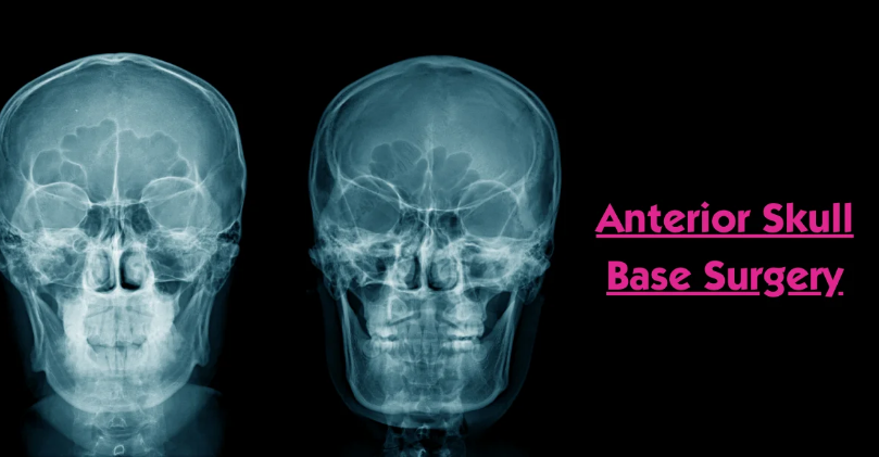
Role of MRI in Surgery of Skull Base: To achieve more ‘precise’ surgery and assure better patient outcomes, MRI is considered one of the most important diagnostic modalities for skull base surgery. It gives very clear, detailed pictures of the brain and other structures surrounding it. Such pictures give surgeons much better perspectives during operations, leading to improved outcomes and safety for the patients. Advanced MRI technology allows the doctor to trace tumors, major blood vessels, and all minute details relevant to successful surgery.
Major advances in the technique of MRI imaging have significantly enhanced its utility in clinical applications. High-resolution scans and functional MRI provide real-time information that surgeons can use to navigate complex areas of the skull base. Such a capability permits precision, which is helpful in reducing risks from such complex procedures.
With the changes that go on within the MRI platform, the impact it has on skull base surgery is equally likely to increase as well. Understanding its use can become an enabling factor for both teams within medicine and patients who undergo surgeries themselves.
Key Takeaways
- MRI provides the necessary images in planning surgery on skull bases.
- Precision and safety are improved with advanced imaging techniques.
- Ongoing technologic development enhances the effectiveness of MRI in surgery.
Imaging Techniques and MRI Technology
In the process of planning and performing skull base surgeries, one of the most vital tools is MRI. Advancement in MRI systems enhances the quality of imaging that enables surgeons to visualize even the most complex structures of the body more clearly. Comparisons among different imaging modalities further provide insights into their effectiveness for surgical preparation.
Advancements in MRI Technology
Over the past two years, there have been considerable improvements in MRI. These include higher magnetic field strengths and better coil design. These improvements offer finer resolution with even quicker scanning times.
Key Recent Advances Include:
- fMRI: Provides a mechanism to evaluate brain function by monitoring flow changes in the brain
- DTI: Delineates white matter tracts, thereby helping in surgical planning.
Such techniques also allow surgeons to get very detailed views of the anatomy of the skull base and, therefore, enhance safety and improve results during actual surgery.
Comparison of Imaging Modalities
There are several imaging modalities available that serve different purposes in skull base surgery. Common ones include MRI, CT scans, and PET scans. All have relative strengths and weaknesses.
Comparison highlights:
- MRI: Excellent for soft tissue contrast, useful in viewing tumors and vascular structures.
- CT Scan: Faster and better for the detection of bony abnormalities.
- PET Scan: It helps in the assessment of the metabolism and function of brain tissues.
The choice of one appropriate imaging modality depends on the particular clinical scenario. In actual surgical practice, most surgeons use a combination of these modalities to comprehend all information relevant to a particular case before undertaking surgery.
Clinical Applications in Skull Base Surgery
Skull base surgery would greatly involve the use of MRI. These provide detailed images that help in the planning, guiding, and assessing of surgeries. Understanding its applications can enhance surgical outcomes.
Preoperative Assessment and Planning
MRI forms part of preoperative assessment and hence gives information on the brain and the surrounding structures, aiding in tumor identification, vascular issues, and some anatomic variations.
Main features attributed to this modality include:
- Tumor characterization: MRI can identify the type of tumor.
- Anatomical mapping: It gives the relation of tumors to critical structures like nerves and blood vessels.
Surgical planning: Surgeons are able to look at particular images to visualize the access route to the target area.
This, in turn, enables surgeons to make appropriate plans and predict the complications of the intervention in advance.
Intraoperative Guidance
MRI offers intraoperative guidance during open surgery. It ensures that the procedure is accurate and complications minimal.
Points to note include:
- iMRI: Some operating theatres have iMRI scanners that provide real-time images during an intervention.
- Tumor resection: Surgeons can verify the complete removal of the tumor.
- Avoiding damage: Real-time imaging views around critical structures limit the number of complications.
This allows for improved operational decisions and the delivery of enhanced safety and effectiveness for the patient.
Postoperative Evaluation
MRI is an important modality during the postoperative phase. It helps confirm the results and any complications after surgery.
A few key factors to look at include:
- Resection evaluation: MRI can verify if the tumor has been completely resected.
- Complication detection: MRI can help find complications such as hemorrhage or fluid collection.
- Follow-up study: Follow-up MRI studies may sometimes be conducted periodically to investigate recurrence.
The purpose of postoperative assessment is to guide further treatment and ensure the best long-term results for the patient.

Also Read :
