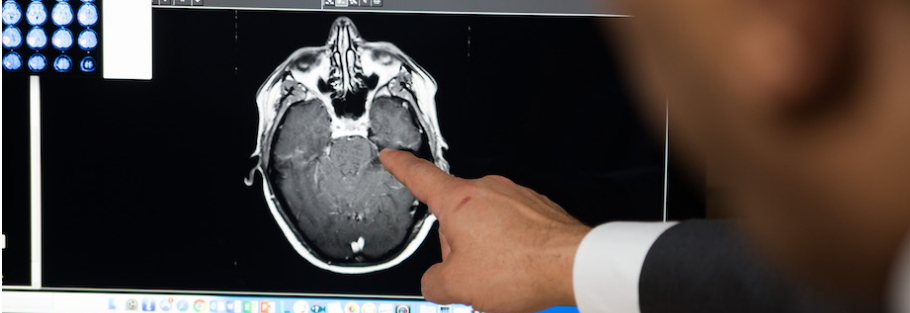
fMRI of the Brain for Neurosurgical Planning in the Surgery of Brain Tumors: Improvement of Surgical Outcome and Safety
Functional MRI is an important modality for neurosurgical planning in the surgery of brain tumors. It helps the surgeon identify the eloquent brain region to safely resect the tumor without compromising vital functions. Advanced imaging by means of fMRI significantly improves surgical outcome.
It is an ideal modality for visualizing the mapping activity of the brain, hence helping the doctor understand which parts of the brain are involved in major activities like talking, movement, and many others. This information becomes critical because tumors may grow into these sensitive areas and, therefore, are very important to know about in advance so that possible complications in surgery can be avoided. Functional MRI is significant in the surgical planning of brain lesions; it is a means toward more precision, facilitating informed decisions.
With the continuous development in technology, functional magnetic resonance imaging is considered an essential modality in neurosurgery. This blog examines how functional MRI is shifting the paradigm in the surgical management of brain tumors and providing improved outcomes for patients.
Key Takeaways
- fMRI aids in identifying key areas within the brain during surgery.
- It enhances safety during tumor removal by guiding surgeons.
- Advanced imaging techniques enhance overall surgical planning and outcomes.
Basics of Functional MRI
fMRI plays a vital role in the surgery of brain tumors by mapping brain activity for the surgeon. This section shall cover the principles of fMRI and its comparison with other imaging modalities.
Principles of fMRI
fMRI relies on the principle of detecting altered blood flow in the brain. This technique exploits the fact that active areas of the brain require more oxygen. Thus, when neurons are active, they consume oxygen, and as a result of this, blood flow increases in that area.
This signal is referred to as the BOLD, or Blood Oxygen Level Dependent, signal. Images obtained through fMRI scan depict this blood flow to the doctor, hence allowing him to identify which parts of the brain are responsible for certain functions, including movement or speech.
FMRI can achieve good spatial resolution in the measurement of brain activity. It captures the dynamic changes in activity of the brain over time. This constitutes its efficacy in studying the function of the brain during tasks or rest.
fMRI compared to Other Imaging Modalities
fMRI is unlike other imaging modalities, like CT and PET scans. For instance, in as much as structural images from CT are of very good quality, they don’t show images of the activities of the brain. On the contrary, PET scans can show an image of brain function. However, most of them use radioactive tracers.
Key Differences:
- fMRI: It basically measures changes in blood flow to create a map about the activity of the brain. It is non-invasive and doesn’t involve the use of radiation.
- CT Scan: It’s good for structural imaging. The idea involves the use of X-rays. This gives a very detailed picture about the anatomy of the brain.
- PET Scan: Provides functional data but uses radioactivity. Not as frequently used for routine brain examinations.
These differences make fMRI especially useful during neurosurgical planning. Since the functioning brain is known, surgeons are more capable of avoiding those areas that control vital functions.
Application in Brain Tumor Surgery
fMRI has become an essential tool in maximizing the surgical benefit in patients with brain tumors. It helps determine an optimal surgical strategy, is useful in making appropriate intraoperative decisions in real time, and is helpful in risk assessment and determinants of patient’s outcome.
Preoperative Planning
fMRI during preoperative planning helps to identify several key brain regions. Surgeons use fMRI to identify which parts of the brain light up when their patients execute certain functional tasks, like speaking, swinging arms and legs, and feeling sensations.
By having this information, they are able to plan routes of tumor removal that are safest. Surgeons will be able to minimize their damage to brain functions by not going near these important areas.
A good example is locating the primary motor cortex using fMRI before surgery. This insight provides a great deal of chance for a successful operation with fewer complications.
Intraoperative Decision Making
Intra-operatively, fMRI can be combined with other forms of imaging. Immediate access to fMRI data can be obtained by surgeons to make decisions about the need for adjustments in strategy during the operation.
They can modify their strategy in response to unexpected findings according to the functional maps. They will often vary their path if they hit an area responsible for speech or motor skills.
This real-time use of fMRI helps ensure that the surgical team preserves vital brain functions, leading to better patient outcomes.
Risk Assessment and Patient Outcome
- fMRI also helps in risk assessment and reviewing the expected patient outcome. With an understanding of functional anatomy, healthcare providers can assess complications.
- Indeed, it was observed that the postoperative care of patients became increasingly individualized once fMRI data were taken into consideration. Such information helps guide rehabilitation efforts and follow-up treatment.
- Eventually, this could be translated to shorter recovery times and an improved quality of life following fMRI-guided surgical resections of brain tumors.

Also Read :
