
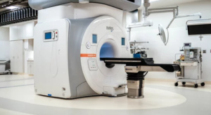
MRI in Pediatric Neurosurgery: Accommodating Special Challenges and Considerations The role of MRI in pediatric neurosurgery includes the provision of clear, sharp images of the brain of a child. To date, the unique challenges in pediatric MRI studies have been brought about by the size of the child, sedation, and variability in cooperation-a factor that may affect image quality. Understanding these challenges is important for any health expert in order to achieve correct diagnosis and effective treatment planning.
Pediatric neurosurgery needs the most correct imaging for a lot of conditions in this area. In recent times, with the advancement of MRI technology, doctors can see and manage problems such as tumors, congenital anomalies, and traumatic injuries more elaborately. These technologies, however, need adapted methods for small patients: one has to make provision in respect of safety and efficacy.
By acknowledging these operational difficulties in pediatric MRI, health care providers are able to create plans and programs which better the patient experience. The ability to address these issues leads to higher success rates of imaging, which in turn enhances outcomes for children undergoing neurosurgical intervention.
Key Points
- MRI is an important modality in the diagnosis and management of brain disorders in children.
- Smaller size and the need for sedation create special challenges.
- Special approaches can help optimize imaging quality and patient care.
Clinical Applications of MRI in Pediatric Neurosurgery
MRI also plays a central role in pediatric neurosurgery by providing maximum details in diagnosis and treatment planning. Applications range from the localization of brain tumors to vascular anomalies.
Localization of Brain Tumors
MRI is of prime importance in the localization of brain tumors in children. It provides high-resolution images that can be used to differentiate tumor types. This information is paramount for the sake of determining the best modality of treatment.
It helps in visualizing the exact tumor size, location, and relationship to surrounding brain structures. Sometimes, it can even visualize features like edema and enhancement patterns. These are very useful in planning safer and more effective operations.
Assessment of Congenital Malformations
MRI is a very useful method of assessment of congenital malformations in pediatric patients. It facilitates detailed imaging of the development of brain structures. Such images may be used to visualize conditions like Chiari malformation or holoprosencephaly.
The ability to visualize soft tissue contrast gives MRI an edge over all other imaging modalities and assists the physician in the detection of abnormalities in brain development and maturation. This becomes very crucial in deciding on the kind of interventions and therapies.
Vascular Anomalies Evaluation
Vascular anomaly evaluation in pediatric neurosurgery is performed using MRI. AVMs are a few of the conditions detected quite easily through MRI. Imaging assists in assessing the flow, as well as structural information within the vascular system.
MRI angiography is very useful to see the blood vessels. It helps in surgical planning and calculation of risks with vascular conditions. Detailed assessment thus provides the safest therapeutic option for a young patient.
Guiding Epilepsy Surgery
MRI has been very instrumental in guiding epilepsy surgery in children. MRI is used for localizing the zone epileptogenetic which generates seizures. This exact localization is crucial for good surgical results.
Advanced techniques can allow MRI to detect abnormalities in the brain tissue not evident through any other means. This knowledge of these areas helps surgeons zero in on the right location during the operations. The result would be a greater chance to reduce seizure frequency or achieve seizure freedom.
Operational Challenges and Solutions
MRI of pediatric neurosurgery has special operational difficulties owing to the particular needs of children. Overcoming these difficulties is crucial in acquiring high-quality images while maintaining patient comfort and ensuring safety.
Sedation and Anesthesia: Considerations
Most children must undergo sedation when undergoing MRI scans in order to remain still for clear images. Adequate assessment by a competent professional is important in ascertaining what type of sedation will be safest concerning the child’s age, weight, and previous medical history.
Commonly, options of sedation include:
- Nitrous Oxide : Mild sedation, rapid recovery
- Oral Sedatives: Useful in patients with older children who swallow pills
- IV Sedation: Younger or more anxious patient deeper sedation possible
Anaesthesia teams need to be specifically trained in pediatric care; close monitoring is necessary to minimize complications associated with sedation. Recovery times after the scan may be variable so needs planning.
Patient Movement and Image Artefact Reduction
This may result in unclear images due to the artefacts produced by the movement. Younger children might find this very challenging.
To minimize movements, personnel can:
- Engage in Play Therapy: This is a way of persuading the child to relax before going into the scan.
- Perform Visual Distractions: Watching movies or playing with toys is usually favored by them and will distract the child from what is happening.
Discuss the procedure with the parent to make sure a comfortable and supportive atmosphere is present.
Padding and restraints can also be fitted on the MRI machine to keep the child in a stationary position during the scan. Such methods will improve image quality while reducing repeat scans.
Magnetic Field Risks and Pediatric Safety Protocols
The use of MRI is not without safety concerns. Children may not understand the dangers of a powerful magnetic field.
Organizations should thus have tight safety protocols in place that
- Screening: this is the assurance that all personnel check for metallic objects prior to entering the MRI area
- Education: explaining precisely what is going to happen to both child and care
- Environment: providing a child-friendly waiting area, reassurance for the patient while distraction from anxiety.
Proper training in the procedures of emergency cases is also needed. The personnel need to be prepared with safety incidents involving a magnetic field. These are protocols to be followed so that the MRI experience can be safe and effective for pediatric patients.
Also Read :