The Role of Magnetic Resonance Imaging in Neurosurgical Trauma Care: Improvement in Diagnosis and Treatment Outcomes MRI is among the very cornerstones in the management of neurosurgical trauma patients. This neuroimaging technique provides great detail of both the brain and the spinal cord, thus greatly helping the clinician in diagnosing injury and effectively planning treatment. This novel imaging technique enables insight into material that is invaluable to emergency informed decisions and to the ongoing informed care of the patient.
Further elucidation of how MRI works enhances the management of traumatic brain injuries. Besides locating the extent of damage, it helps neurosurgeons during operations. The ability to visualize the injury accurately is a prime key that enables improvement in patient outcomes and minimizes risks.
Advances in the modality continue to expand the role of MRI in trauma. With its clear, accurate pictures, MRI continues to play a vital role in medical diagnosis, especially in conditions that require immediate neurosurgery.
Key Points
- MRI provides necessary images of brain and spinal injuries.
- It allows for surgical planning and management of the patient.
- Advances continue to further enhance its utility in trauma care.
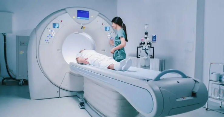
Basic Principles of MRI Technology in Neurotrauma
MRI is very important in neurotrauma because it provides minute details of both the brain and the spinal cord. Diagnosis and treatment will be effective only when there is an understanding of the physics of the technology and safety considerations while using it.
Physics of MRI and Image Acquisition
MRI works by producing images of the human body using a strong magnet combined with radio waves. First, the machine produces a magnetic field. It aligns the hydrogen atoms in the body, mostly situated in water and fat.
When radio waves are applied to the atoms, they emit signals. The MRI will detect these signals and convert them into images. The images can show a number of injuries of the brain, tumors, or any other abnormalities. Some of the important terms in the process are:
- Strength of the magnetic field: It is usually measured in Tesla
- Radiofrequency pulses: These are used for exciting the atoms.
Different imaging techniques, such as T1 and T2-weighted images, serve to emphasize different tissues. These provide a vivid view of brain conditions for the doctors.
Safety and Precautionary Measures in MRI Use
Safety during MRI scans is an area of great concern. The strong magnetic field exerts an attraction force on metallic objects, thus posing a danger. Patients are, therefore, required to report any metal implants and devices in their body.
Before undergoing an MRI, patients normally have to go through this checklist:
- Metal implants: Are there any?
- Claustrophobia: Would this be a problem?
- Contrast agents: Does further imaging with these agents become necessary?
The MRI environment is made as “harm-reduced” as possible. Technologists are present and monitor the patient during the scanning. They also give ear protection to help protect the ears from the loud machine noises.
These precautions mean that MRI continues to be a safe, yet effective, modality in neurotrauma assessment.
Clinical Applications of MRI in Neurosurgical Trauma
Over the years, MRI has become the central tool in diagnosing and managing neurosurgical traumas by establishing the extent of the injury, surgical planning, and follow-up. Details related to these major uses have been discussed in the subsequent paragraphs.
Assessment of Acute Brain Injuries
Acute traumatic brain injuries are not easily detected; therefore, the need for MRI to outline the structural anatomy of the brain is of particular significance. In this case, imaging will contribute to the diagnosis of diseases such as contusions, hemorrhages, and edema.
The following are the key MRI findings in TBI:
- Contusions: Blood collects in the brain tissue.
- Hemorrhages: The vessels may burst and bleed.
- Edema: There is swelling due to injury.
With the use of MRI, the clinicians are able to assess the extent of the above injuries. This clarity helps them determine the immediate course of treatment. It is very important to identify the slight changes in the brain tissue. It might affect surgical intervention and the care of the patient.
Preoperative Planning and Navigation
Before surgery, MRI aids in the preoperative planning of neurosurgery through the good visualization it offers in brain anatomy. Such detailed imaging guides neurosurgeons in preparation for surgery.
Some benefits that MRI provides regarding preoperative planning include:
- Lesion localization
- Mapping of essential brain function
- Assessment of tissue health surrounding the lesion
This information serves to guide surgeons around critical areas. It ensures that surgical strategies can be both safer and more effective. MRI data is integrated into various navigation systems. This assures accuracy during surgical intervention.
Monitoring and Postoperative Evaluation
MRI after surgery is necessary and is crucial for post-surgical follow up. MRI essentially details the extent of recovery of the brain. This imaging technique may also detail the complications such as infection or new hemorrhage.
MRI’s of a patient after surgery may include:
- Analysis of re-bleeding
- Checking the healing at the site of surgery
- Functional improvement.
Regular scans guide follow-up care. They allow changes in rehabilitation strategies based on current data. This way, continuous assessment enables the best recovery possible for the patient. MRI stands as a substantial tool in the whole trauma care process.

Also Read :