
Neurosurgical MRI Artifacts: Understanding Identification and Minimizing Imaging Challenges In neurosurgery, however, MRI technology has become an essential modality for amazingly detailed pictures of the brain and other structures surrounding it. Understanding MRI artifact is important since artifacts are conditions that may lead to misinterpretation and alter surgical decisions. The doctor thereby addresses these issues to improve the outcomes of patients undergoing neurosurgery.
Neurologists and radiologists should be aware of commonly occurring MRI artifacts due to motion, magnetic field inhomogeneities, and limitations of equipment; this will provide them with appropriate ability concerning image interpretation and the capability of making appropriate treatment decisions. Knowing how to identify and solve these artifacts can facilitate further quality care of patients.
Understanding various MRI artifacts helps healthcare professionals navigate the intricacies of neurosurgery. This article will demystify those critical aspects of MRI imaging that guarantee accuracy in the operation theater and beyond.
Key Takeaways
- MRI is a necessity for accurate brain imaging when it comes to neurosurgery.
- Being able to identify and correct artifacts can avoid incorrect interpretation of the images.
- Understanding improves surgical outcomes for patients.
Basics of MRI in Neurosurgery
The basic role of the MRI modality in its extensive function within neurosurgery is to deliver differential highly contrasted images of the brain and other structures of the head. Knowledge of its principles and applications is thereby useful in the process of planning a surgical intervention.
Basic Principles of MRI Technology
MRI scans make use of powerful magnetic fields and radio waves to create images of organs and tissues inside the body. The machine sends out a magnetic field that aligns protons within the body. When this radio frequency pulse is turned on, it will knock the protons out of alignment. As the protons return to their previous position, they emit signals captured by the scanner.
These are the signals that are processed to produce a very detailed image. MRI is useful in well-contrasting various types of tissues, especially in visualizing brain structures. The lack of ionizing radiation makes MRI safer than CT for repeated examination.
Role of MRI in Neurosurgical Planning
Neurosurgery: MRI enables the surgeon to locate a tumor along with other vital areas, such as blood vessels. It helps surgeons decide about the size, shape, and location of a tumor before entering the operation theater. These details help them to plan the best surgery.
Besides this, MRI is able to depict the proximity of a tumor to structures of the brain that are very vital. This kind of information allows for less invasive surgery with fewer risks of complications. It also aids in assessing conditions such as traumatic brain injuries and abnormalities.
Types of MRI Sequences Used in Neurosurgery
There are various kinds of MRI sequences, which fall under different categories depending on the type of image required. The commonly used include:
- T1-weighted images: Best for anatomical detail. They depict fat as bright and are useful during the examination of tumors and their texture.
- T2-weighted images: Useful to review fluid and edema. They help identify abnormalities in cerebral contusions or lesions.
- FLAIR: This is a sequence that suppresses the signal of fluids, thus being effective in finding lesions near the cerebrospinal fluid.
- Diffusion Weighted Imaging-DWI: This detects areas of restricted water movement, which is effective in the diagnosis of acute strokes.
These sequences provide broad insights that are necessary for proper neurosurgical planning and intervention.
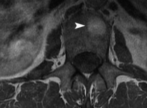
MR Image Artifacts: Detection and Solutions
MRI images may reveal a variety of artifacts, each having an effect on the quality of neurosurgical imaging. The detection of these artifacts with the application of workable solutions is thought to be important for accurate diagnosis and treatment planning.
Common Artifacts in Neurosurgical Imaging
A variety of artifacts commonly arise in MRI scans. There are motion artifacts that result when a patient has moved during the scan. These usually give out-of-focus images.
Another common artifact involves susceptibility artifacts. Magnetic field inhomogeneities generate these, especially around air-tissue interfaces, such as the sinuses. They can cause distortion in the images.
Another form of chemical shift artifacts is when fat and water resonate at slightly different frequencies, misregistering tissues and obscuring significant structures within the brain.
Sources of MRI Artifacts
Artifacts can be sourced from various areas. Of these, the most critical is motion of the patient. Any form of motion, whether breathing or shifting, will produce image distortions.
Artifacts can also be introduced due to equipment-related factors. Variability of magnetic field strength or coil sensitivity may result in incorrect signal capture.
Other environmental elements, such as electronic devices within close proximity, interfere with the MRI machine and produce additional noise. Additional noise degradation leads to worse image quality.
Artifact Reduction Strategies
Several techniques can be adopted to reduce the appearance of artifacts in MRI images. The most significant preparation involves the briefing of the patient on the importance of remaining still during the test. Cushions and straps can be used to comfort them but also keep them in place.
The next step involves manipulating the imaging parameters to assist in reducing the artifacts. Lowering TE can reduce motion artifacts. Selection of appropriate pulse sequence can help in minimizing susceptibility artifact.
Additionally, proper selection of coil can enhance the quality of the image. Use of a dedicated head coil while doing a brain can reduce noise and hence provide clear images.
Correction Techniques for Artifacts
A variety of techniques can correct MRI artifacts after occurrence. For instance, motion correction algorithms can be applied to enhance images degraded by motion of patients. These algorithms manipulate the images according to known patterns of movement.
Other techniques include the usage of fat suppression techniques. These techniques nullify the chemical shift artifacts, hence providing sharper images of brain tissues.
Advanced post-processing tools can also be applied. They analyze the data of images to identify different types of artifacts and further correct them, hence enhancing the overall image quality. The use of such tools can make a huge difference in neurosurgical images.
Also Read :