Neurosurgical Approach for the Treatment of Brain Aneurysm
A brain aneurysm is a bulging or ballooning in the blood vessel of the brain. After some time, the wall that has weakened in the blood vessel will finally rupture and is likely to cause severe complications later on in life, that may be life-threatening and include stroke, brain damage, or death. Not all aneurysms rupture; however, when they do, the results can often be disastrous. Fortunately, neurosurgical intervention has ensured effective treatments for brain aneurysms, allowing the majority of patients to make a full recovery and return to daily activities.
The following paper discusses neurosurgical approaches to the treatment of brain aneurysms by touching on various types of aneurysms, diagnosis, and surgical techniques. We also discuss the risks and follow-up recovery processes and the importance of early detection to prevent rupture.
1. Understanding Brain Aneurysms
What is a Brain Aneurysm?
A brain aneurysm results from a weakness in one spot of the wall of a blood vessel that bulges outward or balloons out. This causes a pocket of blood to form which can press on surrounding brain tissue and nerves. Brain aneurysms can be divided into classes according to shape, size, and part of the brain involved.
There are two basic types of brain aneurysms:
Saccular Aneurysms (Berry Aneurysms): These are the most common types of brain aneurysms and have a rounded or berry-like shape. They often arise at the junction of blood vessels and can increase in size over time.
Fusiform Aneurysms: These aneurysms involve a dilation of the blood vessel and do not have a clear “neck” like saccular aneurysms. They are less common but can be more difficult to treat.
Aneurysms are more likely to happen in the arteries that form a circle around the base of the brain, a complex junction. Although many aneurysms do not have symptoms and can be small, they may turn dangerous when they increase in size or rupture.
Risk Factors for Brain Aneurysm:
Several factors contribute to the chance of a brain aneurysm forming or rupturing an existing one:
- High Blood Pressure (Hypertension): High blood pressure thins the walls of the blood vessels, thereby increasing the risk for rupture of the aneurysm.
- Genetic Predisposition: Genetic predisposition due to a history of brain aneurysms in families may be related to genetic origin.
- Smoking: Smoking destroys the blood vessels, accelerating the formation and rupture of an aneurysm.
Age and Gender: Aneurysms are more common in individuals between the ages of 35 and 60 years and are more likely to occur in women.
Other Medical Conditions: Conditions like polycystic kidney disease, connective tissue disorders, and brain arteriovenous malformations (AVMs) may increase the risk of developing an aneurysm.
2. Symptoms of Brain Aneurysms
Most brain aneurysms do not cause symptoms until they rupture. In many cases, individuals with unruptured aneurysms may not know they have one. However, larger aneurysms or those that press on surrounding brain tissue may cause signs of a problem, including:
Headaches: A sudden severe headache can be a warning symptom, especially if unlike other headaches. Vision disturbances may include double vision, one side blurred or visual loss if the aneurysm affects nerves that control the eyes. Nausea and vomiting are common complaints with a ruptured aneurysm due to pressure on the brain. Seizures: Brain aneurysms can cause seizures especially if it is large or increasing in size.
Numbness or Weakness: When the aneurysm involves a particular area of the brain, it may lead to weakness or numbness on one side of the body. Neck Pain or Stiffness: Pressure from the aneurysm can cause pain or stiffness in the neck.
If an aneurysm ruptures, the patient may experience a “thunderclap headache,” described as the worst headache, with tremendous sudden intensity. In such cases, a visit to a doctor is an absolute emergency.
3. Diagnosis of Brain Aneurysms
It is very important to detect the brain aneurysms as early as possible to avoid their rupture. Most of the patients are diagnosed while going for some routine imaging studies. Various diagnostic modalities include:
CT Scan (Computed Tomography)
A CT scan can find a brain aneurysm fairly quickly. If the aneurysm has ruptured, then the test can find blood in the brain, which means there was a hemorrhagic stroke. A CT scan may be ordered to plan for further testing.
MRI (Magnetic Resonance Imaging)
An MRI gives detailed pictures of the brain and blood vessels. Doctors can see the size and location of an aneurysm. A type of MRI is called MRA, or Magnetic Resonance Angiography. MRA uses magnetic resonance technology to see only the blood vessels.
Cerebral Angiography
Cerebral angiography is considered the gold standard in the visualization of cerebral aneurysms. In this, a catheter is introduced into the vessels, after which contrast dye is administered. X-rays are then taken, which show details of the vessels in the brain. This is usually indicated for assessing the size, shape, and place of the aneurysm pre-operatively.
4. Brain Aneurysm Treatment Options
When a brain aneurysm is diagnosed, treatment depends upon size and location of the aneurysm, besides the general condition of the patient’s health. The two available main methods for treatment are observation and surgery.
1. Observation
For small, unruptured aneurysms that are asymptomatic, a neurosurgeon may advise a “wait-and-see” approach. The patient may be followed with periodic imaging to ensure the aneurysm is not growing or changing. This approach is typically reserved for patients who are low-risk for rupture and whose aneurysms are asymptomatic.
2. Neurosurgery for Brain Aneurysms
Larger aneurysms and symptomatic or those which have a chance of rupture warrant surgical interventions. Neurosurgical treatments for the conditions of brain aneurysm primarily involve clipping and coiling.
Surgical Clipping: Conventional treatment of the aneurysm of the brain includes surgical clipping. The surgical operation involves the craniotomy of the skull to approach the aneurysm. After the detection of the aneurysm, a small metallic clip is being placed around the neck of the aneurysm to hinder blood flow within, thus sealing the aneurysm off. This will in turn prevent it from breaking and bleeding sometime in the future.
Clipping works best for aneurysms appearing on the surface of the brain and is usually performed when the aneurysm is large or positioned in a manner that is very difficult to get to with other means. However, it is an invasive procedure and thus needs a longer period of recovery.
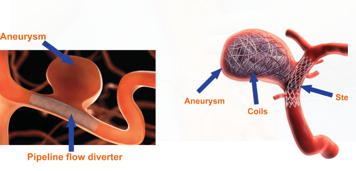
Endovascular Coiling: This is a much less invasive procedure in which, through an artery in the groin or the wrist, a catheter is inserted and then advanced to the site of the aneurysm in the brain. Small platinum coils are then introduced through the catheter into the aneurysm, allowing the formation of a blood clot that effectively seals off the bulging blood vessel.
Coiling is often preferred for aneurysms that are deeper in the brain or in areas hard to reach. It offers a quicker recovery time and less risk of infection compared to surgical clipping. However, coiling may not be suitable for all aneurysms, especially larger ones.
3. Flow Diverter Stents:
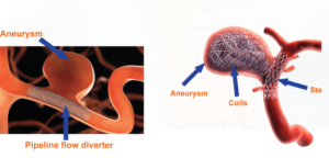
The large or complex aneurysms are treated in some cases with flow diverter stents. The stent is placed inside the blood vessel to reroute the flow of blood past the aneurysm, allowing the blood vessel to heal and the aneurysm to stop continuing to expand.
5. Risks and Complications of Brain Aneurysm Surgery
Neurosurgery for brain aneurysms saves lives, but it isn’t entirely free of risks. Some possible complications include
Stroke: The possibility of stroke, especially in cases of large-sized aneurysms, it might affect the normal anatomy of the patient because disrupting the flow of blood.
Infection: Because it is a surgical approach, there are possible infections that might occur around the area where the incision has been made.
Seizure: Seizures occur infrequently as they are caused due to brain surgery.
Re-bleeding: This hardly ever occurs because once treated, a previously ruptured aneurysm re-bleeds or forms another aneurysm.
Cognitive Changes: Depending on the site of the aneurysm, surgery may result in temporary or permanent cognitive or neurological changes. 6. Recovery and Outlook
The duration of recovery following surgery for a brain aneurysm depends on the type of surgery that has been performed and the individual patient’s health. Patients who have undergone a craniotomy may take several weeks to recover completely, while coiling or flow diversion patients usually have less recovery time.
Tips for Recovery after Surgery
Rest: Rest is necessary in the initial phases of recovery, avoiding heavy work.
Follow-up Care: Follow-up with regular visits and imaging studies to make sure that the aneurysm has been treated and hasn’t recurred.
Rehabilitation: Some patients require physical therapy or cognitive rehabilitation to fully recover.
Outlook for Brain Aneurysm Patients:
Generally, the prognosis following surgery is usually good for the patients who have undergone treatment for brain aneurysms. The key to a good prognosis is early detection and timely treatment. Many patients who undergo successful treatment can return to their normal activities, though the risk of future aneurysms may still exist.
Conclusion
The neurosurgical approach to treating brain aneurysms is a critical intervention for preventing rupture and preserving brain function. Neurosurgery has advanced to offer effective solutions for patients with brain aneurysms through surgical clipping, endovascular coiling, and flow diverter stents. The key to preventing life-threatening complications and ensuring the best possible outcomes for individuals affected by this condition is early detection, careful evaluation, and timely treatment. If you suspect a brain aneurysm or have risk factors, consult a specialist to assess your situation and explore potential treatment options.

Also Read :
- Key Neurosurgical Procedures Explained in Simple Terms
- What to Expect During Your First Neurosurgical Consultation
- The Neurosurgical Approach to Treating Chronic Migraines
- The Importance of Neurosurgical Research in Treating Rare Disorders
- MRI Safety Considerations in Neurosurgical Patients with Implants
