Imagine looking inside the human body without an incision. Picture a brain, a heart, or a complex tumor floating in three dimensions right before your eyes. This isn’t science fiction; it’s the power of 3D MRI visualization. We’re moving beyond simple 2D slices to create detailed, interactive models. Demand for these advanced 3D renderings is booming across many fields.
This guide will show you how to take raw MRI data and turn it into a complete 3D model. You’ll learn the practical steps, from understanding your source files to polishing your final visualization. This journey is broken down into simple, easy-to-follow instructions.
Who can benefit from this? Medical professionals, researchers, medical device makers, and students, for starters. The advantages are huge: better diagnosis, clearer surgical plans, easier patient education, and big steps forward in research. You’ll gain a skill that truly transforms how we see and understand human anatomy.
Section 1: Understanding Your MRI Data
What is MRI Data?
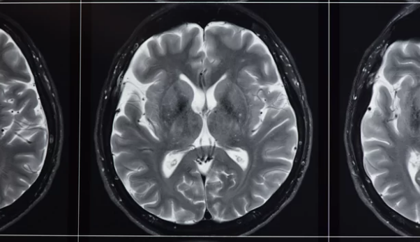
MRI stands for Magnetic Resonance Imaging. It uses strong magnetic fields and radio waves to create detailed images inside the body. Unlike X-rays, MRI doesn’t use radiation. Instead, it measures signals from water molecules in your tissues. Different types of MRI sequences, like T1, T2, and FLAIR, highlight different tissue properties. For example, T1-weighted images show good anatomical detail, while T2-weighted images are better for seeing fluid and swelling. Knowing these differences helps you pick the best scan for your visualization goal.
Data Formats and File Types
The main file format for MRI scans is DICOM (Digital Imaging and Communications in Medicine). Think of a DICOM file as more than just an image. It holds the actual pixel data along with important details like patient information, scan date, imaging settings, and even the hospital where it was taken. This extra information is crucial for accurate 3D reconstruction. While DICOM is king, you might also find NIFTI files, especially in research, which store brain imaging data efficiently.
Data Quality and Considerations
High-quality MRI scans are a must for good 3D models. Issues like patient movement during a scan can cause blurry images, called motion artifacts. Metal implants can also create problems, known as susceptibility artifacts, making parts of the image unreadable. These issues can make it hard to reconstruct accurate 3D shapes. Always check the scan protocols and image quality before you start. A clear, well-scanned image saves a lot of work later on.
Section 2: Software and Tools for 3D Reconstruction
Overview of 3D Visualization Software
Many software options exist for medical 3D imaging. Some are highly specialized, made just for doctors and researchers. Examples include OsiriX, 3D Slicer, and Mimics. Then there are more general 3D modeling programs, like Blender or Autodesk Maya, that can handle medical data with the right plugins. Some newer solutions even offer cloud-based platforms, letting you work from anywhere. Each has its own strengths for creating anatomical visualizations.
Key Features to Look For
When picking software, check for certain must-have features. You’ll need reliable DICOM import capabilities to load your scan data. Strong segmentation tools are vital; these help you separate specific body parts from the rest of the image. Good rendering options are also key. This includes surface rendering, which makes solid mesh models, and volume rendering, which shows the raw data as transparent blocks. Measurement tools and various export options are also super helpful for your project.
Choosing the Right Tool for Your Needs
Picking the best software depends on what you need it for, your budget, and how much you know about 3D work. Are you a beginner or an expert? Do you have money to spend, or do you need free tools? For learning and exploring, try free, open-source software like 3D Slicer. It’s powerful and has a large community to help you out. For professional clinical use, you might need a paid, certified medical imaging package.
Section 3: The Reconstruction Process: Step-by-Step
Importing and Organizing DICOM Data
Your first step is to load the DICOM files into your chosen software. Most programs have an “import DICOM” option. You’ll often see a series of individual image slices. Organize this data logically. Group files by patient name, the date of the scan, or the specific body part scanned. This keeps your workspace tidy and makes it easy to find what you need. A well-organized dataset is the start of a smooth workflow.
Segmentation: Isolating the Anatomy of Interest
Segmentation is where you tell the software which parts of the MRI scan you want to turn into a 3D model. This means outlining or “painting” the specific tissues or structures you’re interested in. You can do this manually, by drawing on each slice. Semi-automatic tools use algorithms to help, like “grow region” or “thresholding.” Fully automatic methods use AI to find structures, though they often need some manual touch-ups. For example, a neurosurgeon might segment a brain tumor to clearly define its borders for surgical planning. Start with simple structures like bone before trying to segment complex, soft tissues.
Creating the 3D Model
Once you’ve segmented your desired anatomy, the software converts that isolated data into a 3D shape. There are two main ways to display this: surface rendering and volume rendering. Surface rendering creates a solid mesh model, like a digital sculpture, often used for 3D printing. Volume rendering shows the original data as semi-transparent blocks, letting you see inside the structure or view multiple tissue types at once. As one biomedical engineer said, “Accurate segmentation isn’t just a step; it’s the foundation for any meaningful clinical diagnosis or research insight from 3D MRI.” The better your segmentation, the more accurate and useful your 3D model will be.
Section 4: Enhancing and Refining Your 3D Visuals
Adjusting Visualization Parameters
Once you have a basic 3D model, you can make it look much better. Play with settings like transparency to see through structures. Use color mapping to highlight different tissues, perhaps making bones white and blood vessels red. Adjust lighting to give your model depth and shadows. Changing the contrast can also make specific features pop out. These tweaks help you show what’s important and make your visuals clearer for others.
Mesh Editing and Smoothing
After creating your 3D model, it might look a bit rough or “jagged” due to the pixel nature of MRI data. You can use smoothing algorithms to soften these edges, making the model look more natural. Sometimes, small holes or imperfections might appear in the mesh. Your software will often have tools to repair these, making your model solid and complete. This post-processing step ensures your 3D visualization is polished and professional.
Adding Labels and Annotations
To make your 3D visuals truly informative, add labels and annotations. You can place text labels directly on anatomical parts, point to specific areas with arrows, or even add measurement indicators. These help explain what viewers are looking at and make complex structures easy to understand. Always use consistent labeling conventions. For instance, always put labels in the same spot, or use the same font, for a clean look across all your models.
Section 5: Applications and Case Studies
Surgical Planning and Simulation
3D MRI models are a game-changer for surgeons. They use these detailed models for pre-operative planning. This lets them see complex anatomy and potential challenges before they even make an incision. For example, a neurosurgeon might use a 3D brain model to carefully plan the safest path to remove a tumor, avoiding critical blood vessels and nerves. This practice leads to more precise surgeries and better patient outcomes.
Medical Education and Patient Communication
Visualizing anatomy in 3D greatly improves learning for students. Instead of flat textbook images, they can rotate and explore realistic models. This also helps doctors explain conditions to patients. Studies show patients understand their health issues much better when doctors use 3D visualizations. A complex heart defect becomes clearer when a patient can see it in 3D, leading to better-informed decisions about their care.
Research and Development
In research, 3D MRI data is a powerful tool. Scientists use it to study how diseases progress over time or to test new treatments without harming patients. It’s also vital for designing and improving medical devices, like implants or surgical tools. As one medical researcher put it, “3D visualization has unlocked new ways to analyze disease and accelerate our understanding of human biology.” It pushes the boundaries of medical science.
Section 6: Exporting and Sharing Your 3D Models
Common Export Formats
After you create your amazing 3D model, you’ll want to share it. Different file formats serve different purposes. STL files are the standard for 3D printing. OBJ and FBX formats are great if you want to use your model in animation or other rendering software. For specific research, especially in neuroimaging, NIFTI files are often preferred because they maintain all the original image data. Pick the format that best fits your next step.
Preparing Models for 3D Printing
If you plan to 3D print your model, there are special steps to take. First, make sure your model is “watertight.” This means it has no holes or gaps in its surface. If it’s not watertight, the 3D printer won’t know what to do. Consider the model’s orientation for printing and ensure its wall thickness is strong enough. You don’t want parts to be too thin and break easily. Always double-check these details before you export for printing.
Best Practices for Sharing
Sharing your 3D models needs some thought. Large files can be hard to send, so consider using cloud storage or specialized sharing platforms. Check that your chosen platform is compatible with the file format you used. Also, be mindful of intellectual property rights and patient privacy. Always get proper consent and remove any identifying patient information before sharing models outside of secure medical networks.
Conclusion
You’ve learned how to transform raw MRI scans into striking 3D visuals. This process, from importing data to detailed segmentation and final rendering, opens up new ways to see inside the human body. The power of these 3D models boosts everything from surgical planning to patient education and medical research.
The future of medical visualization is bright, with AI-assisted reconstruction making the process even faster and more precise. Now, it’s your turn. Start exploring these techniques and tools. The ability to create incredible 3D anatomical models from MRI data is within your reach.
Also Read :