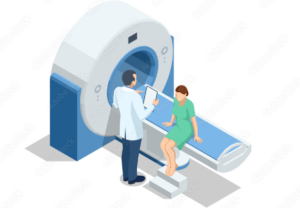
Magnetic Resonance Imaging (MRI) has changed how doctors see inside the human body. It creates detailed pictures of organs and tissues without surgery or harmful radiation. Over time, this technology has evolved beyond simple images to powerful visualization tools that help medical teams understand complex health issues.
The true strength of going from MRI to visualization lies in turning raw data into clear, actionable insights that improve patient care and treatment decisions. This transformation bridges medicine and technology, allowing doctors to plan surgeries, diagnose illnesses, and communicate with patients more effectively. New tools like virtual reality and advanced imaging increase the precision and impact of MRI results.
By combining advanced hardware, software, and medical knowledge, the gap between seeing images and understanding health conditions has narrowed. This growing connection opens new paths for both research and clinical use, making medicine more precise and personalized.
Key Takeways
- MRI technology provides detailed images that reveal internal body structures without surgery.
- Advanced visualization tools turn MRI data into clearer information for patient care.
- Improvements in imaging have led to more accurate diagnosis and treatment planning.
Foundations of MRI Technology
MRI relies on complex physical and technical processes to capture detailed images of the human body. Its effectiveness depends on core scientific principles, advanced hardware, and precise methods for gathering image data. These elements work together to produce high-quality images used in diagnosis and research.
Principles of Magnetic Resonance Imaging
MRI uses strong magnetic fields and radio waves to generate images. It detects signals from hydrogen atoms in water and fat molecules inside the body. These atoms align with the magnetic field, and when radio waves pulse them, they emit signals as they return to their original alignment.
The timing of these signals, called relaxation times, provides contrast between different tissues. This contrast helps distinguish organs, muscles, and other structures. MRI is non-invasive and offers high spatial resolution without harmful radiation.
Advancements in MRI Hardware
Modern MRI machines have improved magnet strength and coil technology. Stronger magnets provide clearer, more detailed scans by increasing signal strength. Advances in coil design allow better detection of signals from targeted body areas.
Computer power has also grown, enabling faster image processing and sharper reconstruction. Hardware improvements focus on enhancing patient comfort with quieter machines and faster scan times. These developments help MRI remain effective in clinical and research settings.
Image Acquisition Techniques
MRI uses various pulse sequences to collect image data. These sequences control the timing and type of radio waves sent and how signals are recorded. Different sequences highlight specific tissue properties or functions.
Techniques like T1-weighted, T2-weighted, and functional MRI capture structural and activity-based information. 3D imaging and faster acquisition methods have become standard. They increase image detail and reduce scan duration, improving diagnostic accuracy.
Integration of MRI with Visualization Tools
MRI data is transformed through advanced techniques that create detailed 3D representations. These visualizations are enhanced by specialized software innovations that improve user interaction and understanding. Effective data processing and interpretation methods help clinicians and researchers make accurate decisions based on complex imaging data.
3D Medical Image Reconstruction
MRI scans produce numerous 2D slices, which must be combined into a 3D model for better spatial understanding. Reconstruction involves aligning these slices carefully to create a volumetric image. This process allows clinicians to view anatomical structures in three dimensions, improving accuracy in diagnosis and treatment planning.
The 3D reconstructions reveal details that are hard to notice in flat images. These models are especially useful in brain imaging, where precise spatial relationships matter. High-resolution and accurate reconstruction techniques enable clinicians to see tissue differences and abnormalities clearly.
Visualization Software Innovations
New software tools have appeared that optimize how MRI data is viewed and manipulated. Many of these programs offer interactive 3D visualization, letting users rotate, zoom, and explore MRI models in real time. Some platforms use virtual reality (VR) to immerse users, making the data easier to understand in complex cases.
These tools often include collaboration features, so doctors and researchers can view images together remotely. They also support various medical imaging formats and integrate with hospital systems. Such innovations improve workflow and patient communication by making images more accessible.
Data Processing and Interpretation
Before visualizing MRI data, raw scans undergo processing to enhance image quality and extract important information. This includes noise reduction, segmentation of tissues, and highlighting regions of interest. These steps prepare the data for clearer visualization and more accurate analysis.
Interpreting MRI visualizations requires algorithms and expert knowledge to identify structures and potential issues. Combining automated data analysis with human expertise helps reduce errors. This layered approach supports clinical decisions, such as surgical planning or monitoring disease progression.
Impact on Diagnosis and Treatment
Medical imaging and visualization technologies have significantly changed how diseases are detected and treated. Advanced tools allow doctors to see detailed images of the body, plan surgeries more precisely, and tailor treatments to individual patients.
Clinical Applications and Case Studies
MRI and other imaging methods help diagnose a wide range of conditions, from brain tumors to joint injuries. For example, 3D MRI scans provide clear views of soft tissues and organs, revealing abnormalities that are hard to detect with standard scans.
In stroke care, fast and accurate imaging guides treatment decisions, improving patient outcomes. Studies show that integrating imaging with data analysis can detect cancers earlier and monitor how well therapies are working.
These clinical uses highlight the growing importance of medical imaging in everyday healthcare, aiding in both diagnosis and ongoing patient care.
Enhanced Surgical Planning
Visualization tools create 3D models from MRI and CT images, allowing surgeons to study anatomy before operations. This helps identify critical structures and plan safer approaches, reducing risks during surgery.
Virtual reality (VR) and augmented reality (AR) platforms let surgeons rehearse procedures or view detailed anatomical maps in real time. This is especially useful for complex surgeries, such as brain or heart operations, where precision is crucial.
Improved surgical planning leads to shorter operation times, fewer complications, and faster recovery for patients.
Personalized Medicine Enabled by Visualization
Medical imaging combined with big data and machine learning supports personalized treatment plans. Doctors can analyze images alongside patient history to recommend therapies that are most likely to succeed.
Visualization helps track changes in tumors or other conditions during treatment, allowing adjustments based on real-time data. This approach focuses on each patient’s unique biology rather than one-size-fits-all methods.
By enabling more accurate diagnoses and targeted treatments, imaging-driven personalization improves care quality and patient outcomes.
Also Read :
