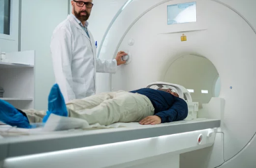
Magnetic Resonance Imaging (MRI) has long been a cornerstone of medical diagnostics, allowing physicians to explore the human body in extraordinary detail without invasive procedures. Over the last few decades, advancements in MRI technology have pushed its applications beyond traditional anatomical imaging into the realm of the mind itself. By offering high-resolution visualizations of brain structures and functions, MRI has opened a new frontier for neuroscience, psychology, and even artificial intelligence research.
In this article, we’ll explore how MRI-based visualizations work, the various techniques used to map brain activity, and the groundbreaking ways these tools are helping scientists unlock the secrets of human thought, emotion, and behavior.
The Evolution of MRI Technology
When MRI technology emerged in the late 1970s, it was primarily designed to provide detailed anatomical images of soft tissues, bones, and organs. Early MRI scans already offered an unprecedented look inside the human body, but the resolution and speed were limited by computing power at the time.
Over the years, refinements in magnetic field strength, coil design, and image processing have dramatically increased the clarity of MRI images. More importantly, innovations such as functional MRI (fMRI) have transformed the machine from a purely anatomical scanner into a powerful tool for studying brain activity in real time.
Today, MRI scanners not only show structural differences in the brain but also capture subtle changes in blood flow, offering insight into how different regions communicate and respond to stimuli.
Functional MRI (fMRI): Mapping Brain Activity in Real Time
The most significant breakthrough in brain imaging has been functional MRI. While traditional MRI focuses on static structures, fMRI measures changes in blood oxygenation levels—a process known as the Blood Oxygen Level Dependent (BOLD) signal.
When a specific brain region becomes active, it demands more oxygen, and the blood supply to that area increases. fMRI detects these variations and translates them into high-resolution, color-coded maps of brain activity.
Researchers use fMRI to:
- Identify which brain areas are engaged during cognitive tasks
- Study emotional processing and decision-making
- Map brain functions before neurosurgery
- Explore differences in brain activity between healthy individuals and patients with neurological disorders
Diffusion Tensor Imaging (DTI): Revealing Brain Connectivity
While fMRI focuses on brain activity, Diffusion Tensor Imaging (DTI) is another MRI-based technique that reveals the brain’s intricate wiring. DTI maps the diffusion of water molecules along white matter tracts, effectively tracing the connections between different brain regions.
This visualization helps researchers understand how neural pathways are organized and how damage to these pathways—through trauma, stroke, or disease—affects brain function. DTI is particularly valuable in studying conditions such as:
- Multiple sclerosis
- Alzheimer’s disease
- Traumatic brain injury
- Autism spectrum disorder
By integrating DTI data with fMRI results, scientists gain a fuller picture of not only where brain activity occurs but also how signals travel through the brain’s communication network.
MRI in Cognitive Neuroscience and Psychology
MRI-based visualizations have revolutionized cognitive neuroscience, allowing researchers to observe the brain in action while subjects perform tasks, experience emotions, or process memories.
For example:
- Memory research: fMRI studies show how the hippocampus engages when recalling past events.
- Language processing: Scans reveal how Broca’s and Wernicke’s areas activate during speech and comprehension.
- Emotional regulation: MRI helps map the interplay between the amygdala and prefrontal cortex in controlling fear responses.
These findings have deepened our understanding of mental health conditions, including depression, anxiety, PTSD, and schizophrenia, enabling the development of targeted therapies.
AI Meets MRI: Predicting Mental States
Recent research combines MRI imaging with artificial intelligence and machine learning to predict mental states and even decode thoughts. By training algorithms on thousands of fMRI scans, AI can detect patterns associated with specific cognitive processes or emotional responses.
This emerging field—sometimes called “mind reading MRI”—has potential applications in:
- Early diagnosis of psychiatric disorders
- Customized treatment plans based on brain activity profiles
- Brain-computer interfaces for individuals with severe disabilities
While the ethical implications are still being debated, the possibilities are both exciting and profound.
Clinical Applications: From Diagnosis to Treatment Planning
MRI-based brain visualizations are invaluable in clinical practice. Neurosurgeons rely on fMRI to avoid critical brain areas during surgery, minimizing the risk of impairing essential functions like speech or movement.
Neurologists use DTI to assess white matter damage after a stroke and guide rehabilitation strategies. Psychiatrists incorporate MRI findings to better understand treatment-resistant depression, using brain imaging to predict whether a patient will respond to medication or therapy.
Moreover, real-time fMRI neurofeedback allows patients to observe and regulate their brain activity consciously, offering promising results in pain management, ADHD, and anxiety reduction.
Ethical and Privacy Considerations
With the ability to visualize thought processes comes the responsibility to handle such data ethically. Brain scans are deeply personal, revealing information about an individual’s mental state, preferences, and potential vulnerabilities.
Key concerns include:
- Informed consent for MRI-based studies
- Data privacy and secure storage of brain images
- Misuse prevention, such as unauthorized “mind reading”
Regulatory frameworks and ethical guidelines are essential to ensure these powerful tools are used only for beneficial purposes.
The Future of MRI-Based Brain Visualization
The next generation of MRI technology promises higher resolution, faster imaging speeds, and more advanced functional mapping. Ultra-high-field MRI scanners (7 Tesla and above) are already providing unprecedented detail in brain imaging.
Future developments may include:
- Portable MRI scanners for bedside brain monitoring
- Combined imaging modalities integrating MRI with EEG or PET scans
- Personalized brain maps for precision medicine
As these innovations progress, MRI will continue to deepen our understanding of the mind—bridging the gap between biology, psychology, and technology.
Conclusion
MRI-based visualizations are more than just medical imaging tools—they are windows into the human mind. From mapping memory pathways to predicting emotional responses, MRI has transformed neuroscience and mental health research.
As technology evolves, the ability to see the brain in action will not only help treat disease but also expand our understanding of human consciousness itself. With careful ethical oversight, the potential of MRI to unlock the mind’s deepest secrets is truly limitless.
If you want, I can also create SEO-optimized meta descriptions, tags, and keywords for this article so it ranks higher in search results. Would you like me to prepare those next?
Also Read :
