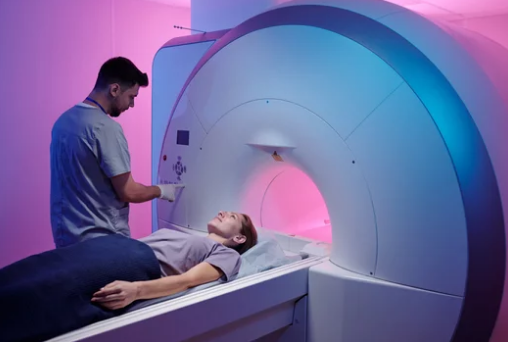
Medical education is evolving rapidly, and one of the most exciting developments is the integration of Magnetic Resonance Imaging (MRI) visualization into the learning process. For medical students, MRI offers more than just a diagnostic tool—it’s a dynamic, interactive way to study anatomy, physiology, and pathology in a living, functioning body.
Through high-resolution imaging and interactive 3D visualization tools, students can now engage with anatomy in ways that go far beyond traditional textbook diagrams or cadaver dissections. This approach not only improves comprehension but also builds clinical thinking skills early in medical training.
Why MRI Visualization is Transforming Medical Education
Traditional anatomy education relies heavily on lectures, cadavers, and static illustrations. While these methods are effective for foundational knowledge, they often fail to show the living, functional aspects of human biology. MRI changes that by providing:
- Detailed soft tissue contrast for clearer anatomical understanding.
- Cross-sectional and 3D views of internal structures.
- Functional imaging to visualize processes like brain activity or blood flow.
- Pathology integration, allowing students to compare normal and diseased anatomy side by side.
Key Benefits of MRI in Medical Student Learning
1. Enhanced Spatial Understanding
MRI slices and reconstructions allow students to view the body in multiple planes—axial, sagittal, and coronal—helping them grasp complex anatomical relationships.
2. Bridging Anatomy and Clinical Practice
Instead of learning anatomy in isolation, students can study MRI scans from real patients, making it easier to connect theoretical knowledge with clinical application.
3. Active, Interactive Learning
When paired with visualization software, MRI datasets can be rotated, zoomed in, or virtually dissected, encouraging exploration and hands-on learning.
4. Early Diagnostic Skills Development
Exposure to MRI interpretation early in training builds confidence and sharpens diagnostic reasoning—skills crucial for future radiologists, surgeons, and general practitioners.
MRI Visualization Tools for Students
Several platforms and technologies now make MRI-based learning accessible to medical students:
- 3D Anatomy Software: Converts MRI data into fully interactive anatomical models.
- Virtual Reality (VR): Immerses students in a virtual body, allowing them to explore structures in real scale.
- Augmented Reality (AR): Overlays MRI scans onto physical models for hybrid learning.
- Cloud-Based MRI Libraries: Provide access to diverse imaging datasets for self-study and research.
Teaching Applications of MRI Visualization
In Anatomy Classes
MRI can supplement or partially replace cadaver work, giving students the chance to explore living anatomy without the limitations of preservation or dissection.
In Clinical Case Studies
Educators can present MRI scans alongside patient histories to teach students how to approach diagnosis systematically.
In Physiology Lessons
Functional MRI (fMRI) brings physiology to life by showing how organs operate in real time—such as neural activation during sensory stimulation.
Challenges to Implementation
Despite its benefits, there are hurdles to widespread adoption:
- Cost: High-quality MRI machines and advanced visualization software can be expensive.
- Training Needs: Students and faculty must learn to navigate imaging software effectively.
- Data Privacy: Patient scans must be anonymized and handled with care to protect confidentiality.
The Future of MRI in Medical Education
As technology advances, MRI visualization will likely become a core component of medical training. We can expect:
- AI-assisted learning platforms that guide students through MRI interpretation.
- Global virtual classrooms where students from different countries can analyze the same MRI dataset in real time.
- Personalized learning modules that adapt MRI-based lessons to each student’s progress.
Conclusion
MRI visualization is revolutionizing how medical students learn anatomy, physiology, and pathology. By offering an interactive, realistic, and clinically relevant perspective, it bridges the gap between theoretical learning and hands-on patient care.
In the coming years, as MRI technology becomes more accessible and affordable, it has the potential to replace passive memorization with active, experiential learning—shaping a new generation of clinicians who can think, diagnose, and treat with precision.
If you want, I can now prepare SEO keywords, meta description, and headline variations so this piece can rank highly in the medical education niche. Would you like me to create those next?
Also Read :