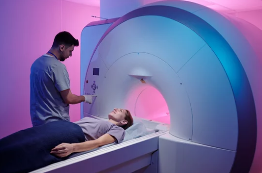
Real-time functional magnetic resonance imaging (fMRI) combined with augmented reality (AR) allows people to see brain activity as it happens. This technology captures detailed images of brain functions and displays them instantly, making it possible to observe how different areas respond during tasks or thoughts. By merging fMRI with AR, brain activity can be visualized live, offering new insights into brain function and behavior.
This fusion gives researchers, doctors, and users a powerful way to understand the brain’s complex processes in a more interactive and accessible way. It supports advancements such as training people to control certain brain functions through neurofeedback. The real-time aspect helps bridge brain data with immediate visual feedback, opening doors for new medical and scientific applications.
Despite its promise, this technology faces challenges like limited speed and the need for precise interpretation. Still, ongoing improvements in hardware and software continue to expand the possibilities of seeing brain activity live in a way that was once impossible.
Key Takeaways
- Brain activity can now be seen live by combining fMRI with augmented reality.
- This technology helps people understand and control specific brain functions.
- Advances are making real-time brain visualization more accurate and useful.
How Real-Time Brain Activity Is Visualized with fMRI and AR
Real-time brain imaging combines detailed brain activity maps with interactive displays, allowing users to see ongoing brain functions as they happen. This process depends on advanced scanning techniques and visual tools to show these changes clearly and quickly.
Overview of Functional MRI Technology
Functional MRI (fMRI) measures brain activity by detecting changes in blood flow. When a brain area is more active, it uses more oxygen, changing the magnetic properties of blood. The fMRI scanner picks up these changes in real time.
The technology uses strong magnetic fields and radio waves to create detailed images. Unlike regular MRI, fMRI captures time-varying signals, revealing which parts of the brain work during specific tasks. The scans are noninvasive and provide high spatial resolution, mapping brain activity with accuracy.
This data is processed rapidly to track brain functions second by second. Real-time fMRI allows researchers and clinicians to observe how brain regions interact dynamically, which is crucial for understanding brain behavior during cognitive tasks or therapy.
Integrating Augmented Reality for Enhanced Visualization
Augmented Reality (AR) overlays digital information onto the real world. When combined with fMRI data, AR can project brain activity visuals onto physical spaces or wearable devices. This integration helps users interact with brain data more naturally.
AR can display 3D brain maps that respond to real-time signals from the fMRI. Users can view changes in brain activity from different angles and zoom levels without needing complex software. This makes interpreting the data easier and faster for neuroscientists and clinicians.
In therapy or research, AR helps visualize the effects of brain stimulation or neurofeedback quickly. It turns abstract data into tangible images, improving communication between patients and medical staff.
Real-Time Data Acquisition and Processing
Real-time fMRI collects brain signals continuously and processes the data instantly. Advanced software decodes the brain’s electrical and metabolic responses right after the scans. This fast processing allows near-immediate visualization of brain states.
The system filters noise and isolates significant changes in blood oxygen levels linked to neural activity. Algorithms convert raw data into visual outputs usable in AR systems. These outputs update every few seconds, reflecting ongoing brain activity without delay.
Combining fMRI with real-time analysis tools, including neurofeedback, users can control or modify brain function consciously. This technique requires precise data handling to ensure accuracy and responsiveness.
Applications in Neuroscience and Clinical Practice
Real-time fMRI with AR supports many neuroscience studies by providing instant views of brain function. Researchers use it to understand cognition, memory, and emotional responses during experiments.
Clinically, it assists in brain mapping before surgery, helping surgeons avoid critical areas. It also aids in monitoring patients with neurological disorders, offering feedback during rehabilitation.
Neurofeedback therapy uses these techniques to help patients regulate brain activity consciously, targeting conditions like anxiety, depression, and PTSD. AR makes this feedback more intuitive by visually representing brain changes clearly during treatment.
These tools improve diagnosis, treatment planning, and patient engagement by making brain data accessible and understandable in real-time.
Challenges and Future Prospects of Real-Time Brain Visualization
Real-time visualization of brain activity using fMRI and AR faces several obstacles. Improving data processing speed, developing user-friendly AR tools, and tailoring methods for individual patients are key areas of focus. These factors shape the next steps in brain imaging technologies.
Technical Hurdles in Real-Time Imaging
Real-time fMRI imaging struggles with temporal resolution. It typically captures brain activity at about 0.5 Hz, limiting the detection of fast brain processes. This slow data acquisition affects how accurately brain function can be monitored during active tasks.
The equipment is also expensive and bulky. High-field MRI machines require large, specialized facilities. Noise and motion artifacts can distort measurements, reducing data quality.
Processing large volumes of data in real time needs powerful computing resources. Algorithms must filter noise while maintaining signal integrity to avoid misleading results. Overcoming these challenges is essential for practical real-time brain visualization.
Improving AR Interfaces for Brain Mapping
Augmented reality can display brain activity directly on a user’s view but requires precise synchronization with imaging data. Current AR devices often lack the spatial accuracy needed for detailed brain mapping.
Designing intuitive interfaces is critical. The system must show meaningful brain activity without overwhelming the user. Visual overlays should highlight relevant areas, such as pain centers or motor regions, in a clear and understandable way.
New AR hardware is becoming lighter and more affordable. However, integrating these devices with fMRI systems needs better wireless data transfer and latency reduction. User testing will help optimize AR tools for clinical and research use.
Potential for Personalized Medicine
Real-time brain visualization holds promise for personalized treatments. It can help doctors track brain changes during therapy and adjust approaches quickly.
For example, neurofeedback using fMRI enables users to learn control over specific brain regions, which may aid mental health disorders. Real-time data could identify treatment responses sooner than traditional methods.
Customizing brain maps for individuals depends on accurate data and efficient processing. As algorithms improve, real-time imaging may guide personalized interventions in areas like rehabilitation and pain management.
Also Read :
