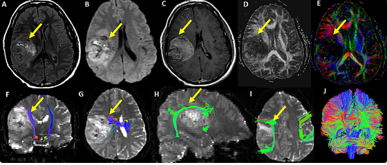
Magnetic Resonance Imaging, or MRI, stands as a cornerstone of modern medicine. It helps doctors see inside the body without cutting it open. This powerful tool provides incredibly detailed pictures of our organs and tissues, guiding diagnoses and treatments daily. But what if we looked past its medical job? Could these complex scans hold a hidden beauty?
The gap between science and art might seem wide, but MRI images truly bridge it. The pictures these machines create are often rich with color and complex shapes. They go beyond simple medical data. Each scan offers a unique view into the human form, showing a visual appeal that feels both scientific and deeply artistic.
This article will explore how MRI scans become stunning visual works. We will look at the science behind these images and unpack their artistic qualities. We will also see how artists and scientists use MRI as a creative tool. Finally, we will consider why seeing medical scans as art can change how we think about both science and ourselves.
The Science Behind the Canvas: Understanding MRI Imaging
How MRI Technology Creates Images
MRI uses strong magnets and radio waves to make pictures of your body. It does not use X-rays or radiation. Inside the machine, a powerful magnetic field lines up the water molecules in your body. Short bursts of radio waves then knock these molecules out of alignment.
When the radio waves turn off, the water molecules relax back into place. As they do, they send out tiny signals. Different tissues, like bone, muscle, or fat, release these signals at different rates. A computer gathers these signals, turning them into visual information.
From Data to Display: The Visualization Process
The raw signals from an MRI scan are just numbers at first. Special computer software takes this data and processes it. This software uses complex math to turn those signals into clear, detailed images. It figures out how strong each signal is and where it came from in the body.
This process helps create contrast, showing clear differences between various body parts. For instance, fluid might look bright white, while bone marrow appears dark. Radiologists and technologists work closely with this process, making sure the images are sharp and accurate for medical study. They often adjust settings to highlight specific features.
The Spectrum of MRI Contrast and Color
Most MRI images you see are black, white, and shades of gray. This is because the signal intensity from different tissues is shown on a grayscale. Bright areas mean strong signals, like from water. Dark areas mean weak signals, like from dense bone.
Sometimes, false colors get added to MRI images during post-processing. This happens often in advanced brain scans or for teaching purposes. These colors are not real but help highlight certain areas or data patterns. When artists use these colorful images, they can create powerful visual pieces that feel both scientific and deeply artistic.
Unveiling the Artistic Elements in MRI Scans
Composition and Form: Nature’s Intricate Designs
Look closely at an MRI image. You will often see surprising beauty in its structure. There is a natural symmetry in our organs, like the two halves of the brain or the chambers of the heart. The patterns of blood vessels or nerve pathways can look like delicate, organic designs.
These images often feel like abstract art, showing forms and curves you never knew existed within you. Think of the swirling patterns of brain folds or the elegant curves of the spine. They show the amazing, complex beauty of biological design. It makes you wonder how such intricate designs could form on their own.
Texture and Detail: A Microscopic Palette
MRI technology captures a rich tapestry of textures. Each tissue type, from soft brain matter to dense muscle, shows up with its own unique visual feel. You can see subtle shifts in brightness that reveal different cell densities or fluid levels. This creates a deeply textured picture.
Specific MRI sequences can highlight even finer details. Some show off nerve fibers, while others focus on tiny blood flow. This detailed information paints a truly unique picture, almost like a microscopic painting. The way light and shadow play on these internal landscapes makes them especially fascinating.
The Emotional Resonance of Medical Imagery
Viewing detailed medical scans can stir deep feelings. Even without knowing a person’s health story, seeing the intricate workings of the human body sparks wonder. It reminds us how delicate and complex life is. You might feel a sense of awe at your own internal world.
These images show our hidden vulnerabilities and strengths. They can make us think about our own existence, our health, and the incredible design of every living thing. This deep connection makes MRI scans more than just diagnostic tools; they become windows into shared human experience.
MRI as Art: Exhibitions and Creative Explorations
The “Brainscape” and Beyond: Artistic Exhibitions of MRI Data
Artists are increasingly turning to MRI data for creative inspiration. Exhibitions featuring “brainscapes” or other body scans have popped up in galleries worldwide. These artists often work with scientists, taking raw MRI data and transforming it into striking visual art.
For example, projects like the “Human Connectome Project” often produce stunning brain maps. These maps inspire artists to explore consciousness and identity through abstract forms. Art installations might project these glowing, colorful brain images onto walls, inviting viewers to walk through an inner landscape. It shows how science can inspire true artistic expression.
Digital Art and Data Visualization: New Frontiers
Digital artists now use advanced software to rework MRI data. They do not just show the scans as they are. Instead, they use algorithms and creative coding to build new visual experiences. This turns complex medical information into something exciting and fresh.
Imagine an interactive artwork where you can “fly through” a brain scan, or a sculpture built from stacked MRI slices. These new art forms help people understand complex science in a new way. They turn raw data into something beautiful and easy to grasp, pushing the boundaries of what art can be.
The Role of Scientific Visualization in Public Engagement
Visually appealing MRI images can make science much more engaging for everyone. When science is shown beautifully, it grabs attention and sparks curiosity. High-quality scientific visualizations help the public understand complex medical ideas. They show the intricate beauty and wonder of the human body.
Institutions can use stunning MRI visuals in educational displays or online. This helps explain how our bodies work or how diseases affect us. Sharing these artistic scientific images encourages a broader interest in science and health. It makes learning both fun and visually captivating.
Expert Perspectives: Scientists and Artists Weigh In
The Interdisciplinary Dialogue: Views from the Medical Field
Doctors and researchers often see the inherent beauty in the MRI images they create. Many radiologists speak of the “art” of image acquisition, where skill and knowledge meet to capture the clearest, most visually striking scans. They know that a well-composed image is not just medically useful but also visually pleasing. This artistic sense can even help them interpret findings more accurately.
Looking at the aesthetic side of MRI images can deepen a medical professional’s appreciation for their own work. It highlights the complex artistry of human anatomy. This perspective sometimes leads to better ways of explaining medical conditions to patients, using the images’ visual power.
The Artist’s Interpretation: Translating Science into Aesthetic Experience
Artists who draw from scientific imagery often feel drawn to MRI scans’ raw beauty. They see patterns, textures, and forms that spark their creative minds. For them, MRI data is not just numbers; it is a blueprint for new artistic expression. They might use color, light, and motion to bring these internal landscapes to life.
These artists often aim to evoke strong feelings in their audience. They want viewers to feel a sense of wonder or even vulnerability. By taking cold, hard data and making it visually compelling, artists help us connect with science on a deeper, more human level. They show us that truth and beauty can live in the same place.
Bridging the Gap: Collaboration and Future Potential
Bringing scientists and artists together creates amazing opportunities. When these different worlds collide, new ideas emerge that neither field might have found alone. Imagine a scientist discovering a new way to visualize complex data thanks to an artist’s fresh perspective. Or an artist finding new inspiration in cutting-edge research.
These partnerships can lead to truly innovative art forms and better ways to explain science. They can spark public interest in health and research, too. The future holds endless possibilities for how MRI, as both science and art, will continue to amaze and inform us all.
Conclusion: The Enduring Beauty of Scientific Vision
MRI technology is primarily a tool for diagnosis, but its artistic qualities are undeniable. It reveals the human body’s incredible detail and intricate design in ways we rarely see. The beauty lies in its ability to capture such complex biological structures with clarity.
More and more, people are appreciating medical images like MRI scans as a form of art. This growing trend helps connect science and culture in new, exciting ways. It encourages us to see the human body not just as a machine, but as a living, breathing masterpiece.
Next time you encounter an MRI image, take a moment to look beyond its clinical use. Appreciate its visual richness. Recognize the scientific wonder it represents. It is a stunning blend of discovery and design, a true testament to the art within science.
Also Read :
