Magnetic Resonance Imaging (MRI) has changed medical care a lot. It gives us amazing views inside the human body. No surgery needed. But MRI data offers more than just health insights. It holds a hidden artistic power. It turns complex body information into beautiful pictures. This article looks at how we make stunning visuals from raw body data. We’ll explore the tools, methods, and creative ways to link science and art.
Medical imaging is now more than just a way to find problems. It has become a source of beauty. MRI shows soft tissues in great detail. This gives a rich palette for seeing the human form in new ways. This journey is not just for good looks. It can help us understand more, teach others, and even make us appreciate our bodies more.
We will show you how to turn MRI datasets into real visual art. We will reveal the core ideas, the software, and the hardware. You will also see how artists and scientists make this happen. Are you a doctor, an artist, a student, or just curious? Prepare to see anatomy in a whole new light.
The Science Behind the Art: Understanding MRI Data Acquisition
Understanding how MRI works helps us see its artistic potential. The data it gathers is the raw material. This section explains the basics of getting MRI data. We will focus on what makes it special for making art.
How MRI Creates Images: From Radio Waves to Pixels
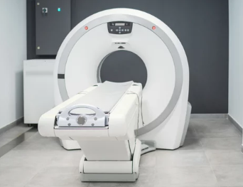
MRI uses strong magnets and radio waves. These excite hydrogen protons in your body. Your body has a lot of water, which means lots of hydrogen atoms. When the radio waves turn off, these protons release energy. The MRI machine detects this energy. It processes these signals to build cross-sectional images. Think of these like slices of your body. Sometimes doctors use contrast agents. These make certain parts of the body show up clearer. This changes how the final image looks.
Types of MRI Sequences and Their Visual Characteristics
Different MRI scans use different “sequences.” These sequences change how tissues appear. For example, T1-weighted scans show fat as bright. T2-weighted scans make water look bright. FLAIR sequences can help spot brain lesions. These differences in how tissues show up are key for artists. They offer varied textures and contrasts. This helps artists pick the best look for their work.
Data Format and Preprocessing: The Raw Material
MRI scanners create data in a special format. DICOM is the most common one. This is like a standard file type for medical images. Before we can visualize this data, we often need to clean it up. This is called preprocessing. Steps like noise reduction remove fuzzy parts. Artifact correction fixes errors from movement. Registration lines up different scans. These steps prepare the data. They make it much clearer for better art.
Translating Data: Software and Techniques for Visualization
Raw MRI scans are just numbers. We need tools and methods to make them visually appealing. This part covers the software and techniques used. We will look at both scientific and artistic methods.
Medical Imaging Software: The Foundation for Visualisation
Many programs help us view MRI data. OsiriX, Horos, 3D Slicer, and Mimics are common choices. These tools let us slice through the body digitally. We can view images from different angles using multi-planar reconstruction (MPR). They also offer initial 3D rendering. This gives a basic 3D view of the scanned area. These programs are the first step in turning data into art.
3D Reconstruction: Building Anatomical Models
Building 3D models from 2D MRI slices is amazing. First, you need to isolate specific body parts. This is called segmentation. You might want to see bones, organs, or blood vessels. You can do this by hand, tracing outlines. Or, automated tools can do it quickly. Once segmented, the software stacks these 2D slices. This creates a full 3D model. It’s like building a sculpture layer by layer.
Artistic Rendering and Manipulation: Beyond Clinical Accuracy
Once you have a 3D model, the fun begins. Artists use many tricks to make visuals stunning. They add color maps to highlight features. Smooth surfaces can make forms look cleaner. Adjusting lighting adds depth and shadow. You can even apply artistic filters. These steps go beyond just showing facts. They let artists create specific feelings or focus on certain features. They truly transform medical data into art.
Case Studies: Where Science Meets Art
MRI data offers amazing chances for art. Here are some real-world examples. They show how MRI images become art. You will see the many ways this blend can happen.
Medical Illustration and Education: Enhancing Understanding
MRI visuals are powerful tools in learning. They appear in textbooks and science papers. Doctors use them to explain complex body parts. A clear MRI-based image helps students learn better than old drawings. For example, animated MRI visuals are great in medical schools. They help explain tricky anatomy ideas. These detailed images make hard concepts easy to grasp.
Art Installations and Digital Art: The Aesthetic Dimension
Some artists use MRI scans as their main material. They explore deep ideas, like what it means to be human. Or they show our inner landscapes. These artists take MRI scans of their own brains. Then they make abstract art from them. Their work makes people think about their own bodies. It shows the hidden beauty inside us all. Their art often appears in galleries and museums.
Personalized Art and Memorials: Unique Expressions of Self
People are starting to make personal art from their own MRI scans. This could be a unique portrait or an abstract piece. It’s a way for people to express themselves uniquely. Some even use MRI data for memorials. Imagine a 3D printed model of a child’s brain tumor. It came right from MRI data. This can be a very powerful and touching reminder. These pieces become truly personal treasures.
The Creative Process: From Scan to Masterpiece
Want to create your own MRI visualizations? This section gives you practical tips. It helps you whether you aim for science or art. Making art from scans can be a rewarding journey.
Selecting the Right Data: Quality and Relevance
Choosing the right MRI scan is important. Look for high resolution. The type of scan sequence also matters. Does it show the body part you care about? Make sure the scan is clear. Also, think about patient privacy. It is very important to use data ethically. Always make sure patient data is anonymous. Get permission if you plan to use it for anything other than medical reasons.
Artistic Interpretation: Choosing a Style and Medium
Now, how do you turn data into art? Think about your style. Do you want bright colors or soft tones? What about the overall look? Plan your composition carefully. Then pick how you will show your art. Will it be a digital print? Maybe a 3D print? Or an animation? These choices help bring your vision to life.
Ethical Considerations and Data Privacy
Working with medical data needs great care. Patient consent is vital. All data must be anonymous. This means removing any patient identifying details. Laws and rules protect this sensitive information. Always follow these guidelines. Get clear permission for any use outside of health care. This protects both the patient and you.
The Future of Anatomical Visualization
The way we visualize anatomy is always changing. New tools and ideas will shape its future. What’s next for seeing our bodies with MRI data?
Advancements in AI and Machine Learning for Visualization
Artificial intelligence (AI) and machine learning are changing things fast. They can automatically segment body parts. This makes the process much faster. AI also makes images look better. It can even create new visualization methods. AI helps make anatomical models more alive and interactive. It saves time and opens new creative doors.
Immersive Technologies: VR and AR in Anatomy
Imagine stepping inside an MRI scan. Virtual Reality (VR) and Augmented Reality (AR) make this possible. These tools create amazing anatomical experiences. They help surgeons plan operations. Medical students can learn in new ways. Even the public can explore human anatomy. These immersive technologies change how we interact with our bodies.
Interdisciplinary Collaboration: Bridging Science and Art
Doctors, scientists, artists, and designers are working together more. This teamwork is key for new ideas. It brings fresh perspectives. This helps both science and art grow. New ways of seeing and understanding emerge. This kind of teamwork pushes the limits of what is possible. It helps us uncover new insights and artistic expressions.
Conclusion: The Enduring Beauty of the Inner Self
MRI data is more than just a diagnostic tool. It is a powerful way to make art. It helps us understand our bodies better. This journey from scan to artwork blends science and creativity.
Recap of Key Takeaways
We have seen how raw MRI data becomes stunning art. It starts with understanding how MRI scans work. Then, special software helps reconstruct 3D models. Finally, artists add their touch. This process shows the amazing mix of scientific knowledge and creative vision.
The Power of Visualization in Appreciation and Innovation
Visualizing anatomy through MRI data does a lot. It helps us appreciate the human body’s complexity. It makes science easier to grasp. And it sparks new forms of art and discovery. The inner workings of our bodies truly hold endless beauty.
Also Read :