MRI scans can seem scary. Knowing what an MRI is, how it works, and why your doctor ordered it really helps patients feel better. This article shows how visual tools and plain talk can make the MRI process clearer. This helps reduce worry and makes the whole experience better for you.
Many patients feel nervous before an MRI. This often happens because they don’t fully get what the test involves. The small space, loud sounds, and how long the scan takes can add to this feeling. Giving patients clear facts, especially with pictures and diagrams, can greatly ease these worries.
This guide will look at many visual resources. Healthcare providers can use them to explain MRI procedures, from basic body parts to the technology itself. By making hard things simple, we can boost patient understanding and happiness.
What is an MRI? A Visual Primer
An MRI uses powerful magnets and radio waves to create detailed pictures inside your body. It helps doctors see soft tissues like organs, muscles, and the brain much better than X-rays. Think of it like taking a very detailed photograph of what’s inside you, without using radiation. This test helps find problems that other scans might miss.
How MRI Machines Work: The Science Behind the Scan
Imagine your body is full of tiny magnets, mostly water. An MRI machine has a big, strong magnet that lines up all these tiny body magnets. Then, it sends out quick radio waves. These waves gently knock the tiny magnets out of line for a moment. When the radio waves turn off, the tiny magnets snap back into place, sending out their own little signals. The MRI machine picks up these signals. It uses a computer to turn them into clear pictures of your insides. This process lets doctors see even very small changes.
What to Expect During an MRI: A Step-by-Step Visual Guide
When you get an MRI, you’ll lie on a comfy table that slides into the machine. The MRI room often looks clean and plain, with the big, round scanner in the center. Before you go in, you might change into a gown. If your doctor needs to see specific things, you might get a special dye injected into a vein. This helps parts of your body show up clearer. During the scan, you’ll hear loud tapping or thumping noises. These sounds are normal and come from the machine working. You’ll often get headphones to help with the noise. The scan can take anywhere from 15 minutes to an hour or more, depending on what your doctor needs to see.
MRI vs. X-ray and CT Scans: When and Why
X-rays use a small amount of radiation to create pictures of bones. CT scans use X-rays too, taking many pictures from different angles to make cross-section views. While great for bones and some bleeding, CT scans also use radiation. MRI, on the other hand, uses no radiation. It’s best for seeing soft tissues. Think of MRI as being able to see a torn ligament in your knee, a small tumor in your brain, or issues with your spinal cord with amazing detail. When doctors need to look closely at organs, muscles, nerves, or the brain, an MRI is often the best choice.
Understanding Your Body with MRI: Visualizing Internal Structures
MRI lets doctors see your body’s inside parts in incredible detail. It’s like having a special camera that shows every tiny piece. This helps them understand what’s going on, whether it’s an injury or a sickness.
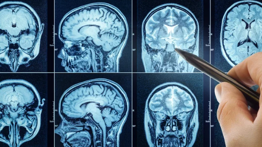
Imaging Your Brain and Nervous System
The MRI is super at looking at your brain and spinal cord. It can show if there’s swelling, a tumor, or even if parts of the brain are not getting enough blood from a stroke. Seeing these detailed pictures helps doctors find nerve problems or conditions that affect thinking and movement. A healthy brain scan looks smooth and even. A scan with problems might show bright or dark spots where they shouldn’t be.
Visualizing Muscles, Ligaments, and Joints
If you hurt your knee or shoulder, an MRI can show the damage. It makes clear pictures of muscles, tendons, and ligaments. These are the soft tissues that connect bones and help you move. For example, an MRI can show if you have a torn ACL in your knee or a rotator cuff injury in your shoulder. It helps doctors decide if you need surgery or another type of treatment. You can see healthy joints next to injured ones, making the damage easy to spot.
The Heart and Blood Vessels: MRI’s Cardiovascular Insights
MRI can check your heart and blood vessels without radiation. It lets doctors see how well your heart pumps blood. They can look at the heart’s chambers and valves. MRI also shows blood flow through your arteries and veins. This is important for finding blockages or other heart problems. Pictures can show the heart beating, helping doctors understand its function very well.
Debunking MRI Myths: Addressing Patient Concerns Visually
Many people have worries about MRI scans. Let’s look at some common fears and how clear visuals can help calm them. Knowing what to expect makes a huge difference.
Claustrophobia and the MRI: Managing the Enclosed Space
Feeling nervous in tight spaces is common for an MRI. Most MRI machines are like a long tube you slide into. But did you know there are also “open” MRI machines? These machines are less enclosed and might be better for you if you get anxious. Doctors might also suggest calming medicines or teach you breathing tricks. Some clinics even show pictures of patients having a scan, looking relaxed. Hearing from others who did it fine can also make you feel braver.
The Noise Factor: What to Expect and Why
The loud knocking sounds during an MRI come from the machine’s strong magnets. These sounds are a normal part of how the images are made. It’s not unlike a washing machine or dryer. You will often get special earplugs or headphones. These block out most of the noise. Some places even let you listen to music through the headphones. Knowing that the noise is just the machine working helps remove some of the fear.
Safety First: Understanding MRI Contraindications
An MRI uses strong magnets. Because of this, some metal objects are not safe to bring near the machine. This includes things inside your body, like certain pacemakers or metal clips from old surgeries. Your doctor or the MRI technologist will ask you a lot of questions about your medical history. They will check for any metal implants you have. This is very important for your safety. They might even show you pictures of what’s safe and what is not safe for the scanner.
Visual Tools in Practice: Enhancing Patient Communication
Clear pictures and videos can make complex medical facts easy to grasp. These visual aids really help patients understand their MRI.
Animated Explainer Videos: Bringing MRI to Life
Short, animated videos can show how an MRI works in a fun way. Imagine seeing tiny protons in your body line up, then get nudged by radio waves. These animations can make the whole process feel less mysterious. They can walk you through the steps of the scan, from entering the room to getting your results. Seeing it happen in a simple video helps you feel more prepared and less confused.
Interactive Apps and Websites: Digital Patient Education
Today, many hospitals have apps or websites where you can learn about your MRI. These digital tools often let you click on parts of a diagram to learn more. You might see a virtual tour of the MRI room. Or you could find quizzes to test your knowledge. This way, you can learn at your own pace, on your own time. These tools put information right in your hands.
Infographics and Diagrams: Simplifying Complex Data
Infographics are like easy-to-read posters with pictures and short facts. They can show how long different scans take or explain how contrast dye moves through your body. Diagrams can also show how the MRI machine takes “slices” or cross-sections of your body to build detailed images. These visual summaries break down complicated facts into small, digestible pieces. This makes them much easier to remember.
Real-Life Imagery: Patient Journey Visuals
Seeing photos of other patients going through an MRI can really help. Pictures of the waiting area, someone getting ready for the scan, or even relaxing inside the machine can make you feel more comfortable. It shows you what the experience is really like. This helps manage expectations and reduces fear of the unknown. Knowing what the room looks like and seeing others go through it can ease your mind.
Expert Insights and Patient Empowerment
Doctors and MRI technologists agree: good communication is key. They often use visual tools to help you understand your MRI.
Doctor and Technologist Perspectives on Visual Aids
Radiologists and MRI technologists often say that when patients see what’s happening, they feel more at ease. “When we show a patient a simple diagram of the MRI machine, their whole face changes,” says one experienced technologist. “They suddenly get it, and their fear drops.” Studies have shown that patients who get clear visual info before a scan are less likely to need the test repeated due to anxiety or movement. This means better pictures and better care for you.
Actionable Tips for Patients Preparing for an MRI
You can take steps to make your MRI go smoothly. Ask your doctor all your questions before the day of the scan. Talk about any fears you have, especially if you get claustrophobic. Wear loose, comfy clothes without zippers or metal. You might even want to bring a friend or family member for support. They can wait for you and help you feel calmer before and after the test.
The Role of Hospitals and Imaging Centers in Visual Communication
Hospitals and imaging centers play a big part in making MRI easy to understand. They can give out special information packets before your appointment. These packets might have diagrams and photos. Many centers now have big screens in waiting rooms showing short videos about MRIs. Training staff to use these visual aids is also important. This ensures everyone gets clear, helpful information.
Conclusion: Clearer Scans, Calmer Patients
Understanding your MRI scan does not have to be hard. Visual tools are powerful ways to make complex medical procedures clear and easy to grasp. By using videos, diagrams, apps, and real-life pictures, healthcare providers can greatly reduce patient anxiety. They can improve your overall experience.
Key Takeaways:
- Visual aids truly help lower stress and improve how patients understand MRI tests.
- Using many types of visuals like animations, charts, and actual photos works best.
- Giving patients clear information helps them feel more in control. This leads to better cooperation during the scan.
- Hospitals and clinics should make visual communication a main part of how they care for patients.
Also Read :