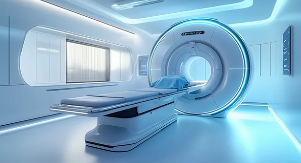
Early detection is critical in the fight against cancer, as it dramatically improves treatment success and patient survival. While conventional imaging methods such as CT scans and standard MRI have been instrumental in diagnosing cancer, advanced MRI techniques are pushing the boundaries of early detection, offering more detailed insights and greater diagnostic accuracy.
This article explores the latest MRI technologies used in early cancer detection, how they work, and why they are changing the landscape of oncology.
Why Early Detection Matters
Cancer is often more treatable when identified at an early stage. Detecting tumors before they grow large or spread allows for less invasive treatments, improved outcomes, and higher survival rates. Advanced MRI techniques enhance the ability to spot cancers at the microscopic or preclinical stage, even before symptoms appear.
Key
Advanced MRI Techniques in Early Cancer Detection
1. Multiparametric MRI (mpMRI)
Multiparametric MRI combines multiple imaging sequences to provide comprehensive information about tissues. Common sequences include:
- T2-weighted imaging – Provides detailed anatomical information.
- Diffusion-weighted imaging (DWI) – Shows water molecule movement, highlighting abnormal tissue.
- Dynamic contrast-enhanced imaging (DCE-MRI) – Tracks blood flow in tissues, revealing tumor angiogenesis (new blood vessel formation).
Applications: Prostate cancer, breast cancer, and liver cancer detection. MpMRI helps differentiate aggressive tumors from benign lesions and guides targeted biopsies.
2. Diffusion-Weighted Imaging (DWI)
DWI measures how water molecules move in tissue. Cancer cells are often densely packed, restricting water diffusion, which DWI can detect.
Benefits:
- Detects small or early-stage tumors.
- Differentiates malignant tissue from inflammation or scar tissue.
- Useful in brain, liver, and breast cancers.
3. Dynamic Contrast-Enhanced MRI (DCE-MRI)
DCE-MRI uses contrast agents to visualize blood flow patterns in tissues. Tumors typically have abnormal vascular networks, which appear as enhanced areas on DCE scans.
Applications:
- Early detection of breast cancer in high-risk women.
- Evaluating liver lesions and identifying hepatocellular carcinoma.
- Monitoring tumor response to therapy.
4. Magnetic Resonance Spectroscopy (MRS)
MRS analyzes the chemical composition of tissues instead of just anatomy. It can detect changes in metabolites that often occur before visible tumors form.
Benefits:
- Differentiates benign from malignant tissue without invasive biopsy.
- Assesses tumor aggressiveness.
- Particularly useful in brain and prostate cancers.
5. Functional MRI (fMRI)
While traditionally used in brain research, fMRI is increasingly applied in oncology to map tumor impact on surrounding functional tissue. For example, in brain tumors, fMRI helps surgeons plan safe resection while preserving critical functions like speech or movement.
6. Whole-Body MRI (WB-MRI)
Whole-body MRI allows scanning of the entire body in a single session, which is especially valuable for detecting metastatic cancer or cancers that are difficult to localize.
Benefits:
- Non-invasive and radiation-free alternative to PET/CT for some cancers.
- Useful in high-risk patients for early cancer surveillance.
- Monitors treatment response and disease progression.
Advantages of Advanced MRI in Early Cancer Detection
- Non-invasive and safe: No ionizing radiation, suitable for repeated scans.
- High sensitivity: Detects small or hidden tumors that other imaging might miss.
- Functional information: Reveals tissue characteristics, blood flow, and metabolic activity.
- Targeted treatment planning: Helps guide biopsies and surgery, reducing unnecessary procedures.
- Monitoring therapy: Detects early changes in tumor behavior before size changes are visible.
Challenges and Limitations
While advanced MRI is powerful, it does have some limitations:
- Cost: Advanced MRI techniques are more expensive than conventional imaging.
- Availability: Not all medical centers have the equipment or expertise.
- Scan time: Some sequences require longer imaging sessions.
- Interpretation complexity: Requires highly trained radiologists to analyze multiparametric or functional images.
- False positives: Highly sensitive scans may detect abnormalities that are not cancer, leading to additional tests.
The Future of MRI in Early Cancer Detection
Emerging technologies promise to make MRI even more effective:
- Artificial Intelligence (AI): AI algorithms help radiologists detect subtle changes and improve diagnostic accuracy.
- Faster scan protocols: Reduced scan times improve patient comfort and accessibility.
- Hybrid imaging: Combining PET with MRI provides both metabolic and structural information for more precise detection.
- Quantitative biomarkers: MRI can now measure tumor characteristics like cellular density and vascularity to predict aggressiveness before clinical symptoms arise.
Conclusion
Advanced MRI techniques are transforming early cancer detection. From multiparametric MRI to magnetic resonance spectroscopy, these tools allow clinicians to see tumors earlier, characterize their aggressiveness, and plan targeted treatments. While not yet universally available, ongoing research and technological improvements are expanding their role in oncology.
If your doctor recommends an advanced MRI, it’s often because it provides the most detailed, non-invasive, and radiation-free view of your tissues, helping detect cancer at the earliest and most treatable stage.
Also Read :
