Magnetic Resonance Imaging (MRI) has long been a cornerstone in medical diagnostics, but recent advancements have expanded its capabilities beyond structural imaging. One such innovation is Functional MRI (fMRI), a technique that measures brain activity and blood flow in real time. While fMRI is widely known for its applications in neuroscience, its potential in cancer research and oncology is gaining attention.
This article explores how fMRI works, its unique advantages, and its emerging role in understanding and treating cancer.
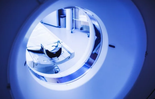
What Is Functional MRI (fMRI)?
Functional MRI is a specialized type of MRI that measures changes in blood oxygen levels (BOLD signals) to map active areas of the brain. Unlike traditional MRI, which produces static images of anatomy, fMRI provides dynamic insights into how tissues function.
In cancer research, fMRI is particularly valuable because it can:
- Assess tumor impact on surrounding tissues
- Study tumor-related brain changes
- Evaluate functional recovery after treatment
- Aid in planning surgical or radiation interventions
How fMRI Works
The principle behind fMRI is based on the fact that active tissues require more oxygen, leading to localized changes in blood flow. Here’s a simplified breakdown:
- Baseline Scan – The MRI records normal blood oxygenation levels.
- Stimulation or Task – The patient performs a task (e.g., speaking, moving a finger) or is scanned during rest to assess spontaneous activity.
- BOLD Signal Detection – fMRI detects small changes in oxygenated vs. deoxygenated blood.
- Data Mapping – Computer algorithms create maps showing which areas of tissue or brain regions are most active.
In oncology, these principles extend beyond the brain to tumor physiology, helping researchers understand tumor metabolism, perfusion, and microenvironment.
Applications of fMRI in Cancer Research
1. Brain Tumor Mapping
fMRI is most widely applied in neuro-oncology. It helps:
- Identify critical functional areas near brain tumors (speech, motor control, vision).
- Plan surgical approaches to remove tumors while preserving essential brain functions.
- Monitor brain reorganization after surgery or radiation therapy.
2. Understanding Tumor Microenvironment
fMRI techniques can assess blood flow and oxygenation in tumors, providing insights into tumor aggressiveness and metabolism. Tumors often create abnormal vascular patterns, which can be detected through advanced fMRI sequences.
3. Evaluating Treatment Response
- Chemotherapy and radiotherapy alter tumor blood flow and metabolism.
- fMRI can non-invasively track these changes over time, offering early indicators of treatment effectiveness before size reduction is visible on conventional imaging.
4. Pain and Symptom Research
Cancer often affects nervous system pathways. fMRI helps researchers study:
- Chronic pain mechanisms in cancer patients
- Neurological side effects of chemotherapy
- Cognitive changes, commonly called “chemo brain”
5. Neuroplasticity Studies
After tumor removal or radiation therapy, fMRI can track brain reorganization, helping rehabilitation specialists design targeted interventions to restore function.
Advantages of fMRI in Cancer Research
- Non-invasive: No radiation, safe for repeated studies
- Functional insights: Provides more than structural imaging, revealing tissue and tumor activity
- Early detection of changes: Detects functional alterations before anatomical changes appear
- Surgical guidance: Protects critical functional areas, reducing complications
- Treatment monitoring: Evaluates therapy effectiveness and guides adjustments
Challenges and Limitations
Despite its potential, fMRI has some limitations:
- Complexity of interpretation: Requires specialized software and expertise
- Motion sensitivity: Patient movement can degrade image quality
- Limited availability: High-end MRI scanners with fMRI capability are not universally available
- Not a standalone diagnostic tool: fMRI complements structural imaging and other functional assessments, but cannot definitively diagnose cancer alone
Future Directions
The role of fMRI in oncology is expanding through:
- Integration with AI: Machine learning algorithms help analyze complex fMRI data for tumor characterization and treatment prediction
- Hybrid imaging techniques: Combining fMRI with PET or diffusion imaging provides structural, metabolic, and functional insights
- Real-time treatment monitoring: Future applications may allow physicians to observe tumor responses during interventions
- Personalized oncology: Functional data from fMRI can help tailor treatments based on tumor behavior and patient-specific brain function
Conclusion
Functional MRI is no longer limited to neuroscience research. In cancer studies, it provides unique insights into tumor physiology, brain function, and treatment response. While it complements rather than replaces traditional imaging, fMRI offers a non-invasive, dynamic perspective that is invaluable for planning therapies, protecting critical functions, and advancing cancer research.
For patients and researchers alike, fMRI represents a powerful tool in the precision oncology toolkit, enabling safer surgeries, better treatment monitoring, and deeper understanding of tumor biology.