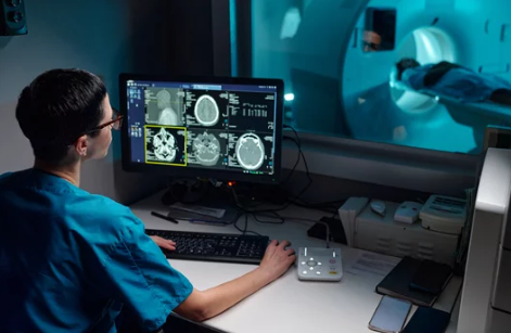
Cancer diagnosis is not just about finding a tumor—it’s also about understanding how far it has spread. This process, known as cancer staging, is essential in determining the most effective treatment plan and predicting patient outcomes. Among the various imaging tools available, Magnetic Resonance Imaging (MRI) stands out for its ability to provide highly detailed images of soft tissues without exposing patients to harmful radiation.
In this article, we’ll explore the vital role MRI plays in staging cancer, how it works, which cancers benefit most, and why doctors rely on it throughout the cancer journey.
What Is Cancer Staging and Why Is It Important?
Staging is the process of identifying how advanced cancer is at the time of diagnosis. Doctors use staging to:
- Determine tumor size and location
- Check if cancer has spread to nearby lymph nodes or distant organs
- Guide treatment choices, such as surgery, chemotherapy, or radiation
- Predict outcomes and survival rates
- Evaluate treatment effectiveness
The most widely used system is the TNM classification:
- T (Tumor): Size and extent of the primary tumor
- N (Nodes): Whether cancer has spread to nearby lymph nodes
- M (Metastasis): Whether cancer has spread to distant parts of the body
MRI plays a critical role in providing the detailed imaging needed for this assessment.
How MRI Helps in Cancer Staging
MRI offers several advantages that make it particularly effective for cancer staging:
1. Detailed Soft Tissue Imaging
MRI provides superior contrast between different types of soft tissue, making it possible to accurately measure tumor size and determine how deeply it has invaded surrounding structures.
2. Multi-Planar Imaging
MRI captures images in multiple planes (axial, coronal, sagittal), giving doctors a three-dimensional view of the tumor and its relationship to nearby organs.
3. Lymph Node Assessment
MRI helps detect enlarged or abnormal lymph nodes, a critical factor in staging cancers like breast, prostate, and rectal cancer.
4. Functional and Advanced Imaging
Techniques such as diffusion-weighted imaging (DWI) and dynamic contrast-enhanced MRI (DCE-MRI) provide information about tumor activity, blood supply, and aggressiveness—data that go beyond simple structural imaging.
5. No Radiation Exposure
Unlike CT or PET scans, MRI does not involve ionizing radiation, making it safer for patients who need repeated scans for staging and follow-up.
Cancers Where MRI Plays a Key Role in Staging
While MRI is useful across many cancers, it is particularly important in staging the following:
1. Brain and Spinal Cord Tumors
MRI is the gold standard for imaging the central nervous system. It reveals the size, location, and spread of tumors within delicate brain and spinal tissues.
2. Breast Cancer
MRI is often used alongside mammography and ultrasound to determine tumor size, check for multiple lesions, and evaluate lymph node involvement.
3. Prostate Cancer
Multiparametric MRI (mpMRI) is essential for staging prostate cancer, allowing doctors to evaluate tumor aggressiveness and whether it has spread beyond the prostate gland.
4. Liver Cancer
MRI provides detailed images of liver tissue, blood vessels, and surrounding structures, helping determine whether tumors are operable or have spread.
5. Rectal and Pelvic Cancers
In rectal cancer, MRI is the preferred method for evaluating how deeply a tumor has invaded the bowel wall and whether lymph nodes or surrounding structures are affected.
6. Gynecological Cancers
For cervical and uterine cancers, MRI helps in assessing tumor spread and guiding surgical planning.
Comparing MRI with Other Imaging Techniques in Staging
To understand MRI’s role better, it’s helpful to compare it with other imaging options:
- CT Scans – Faster and better for staging cancers in the chest, lungs, and abdomen, but less detailed in soft tissues.
- PET Scans – Show metabolic activity of cancer cells and are excellent for detecting metastasis, but often paired with CT or MRI for structural details.
- Ultrasound – Useful in some staging situations (like liver lesions), but not as precise as MRI.
In many cases, MRI is used in combination with CT or PET scans to provide the most accurate staging information.
Advantages of MRI in Cancer Staging
- Excellent for soft tissue detail
- Radiation-free and safe for repeated scans
- Helps with surgical and treatment planning
- Advanced functional imaging provides extra insights
Limitations of MRI in Staging Cancer
Despite its benefits, MRI also has some limitations:
- Higher cost compared to CT scans
- Longer scan times (30–60 minutes), requiring patients to remain still
- Claustrophobia and patient discomfort inside the scanner
- Not ideal for lung or bone cancers, where CT is more effective
- Limited availability in some hospitals or regions
Future of MRI in Cancer Staging
MRI technology is rapidly evolving. Some promising advancements include:
- AI-enhanced imaging to improve accuracy and speed
- Hybrid PET/MRI systems that combine structural and metabolic data in one scan
- Ultra-high-field MRI for sharper and more detailed images
- Biomarker imaging to predict how cancers will respond to specific treatments
These innovations will likely make MRI even more central to cancer staging in the future.
Conclusion
MRI plays a vital role in cancer staging by providing detailed, radiation-free imaging that helps doctors determine the size, spread, and behavior of tumors. From brain and prostate cancers to breast and rectal cancers, MRI is often the go-to tool when precision and soft tissue detail are critical.
While it has some limitations, MRI remains a cornerstone in staging, treatment planning, and ongoing monitoring—making it an invaluable ally in the fight against cancer.
Also Read :
