Magnetic Resonance Imaging (MRI) is a powerful, non-invasive tool in cancer detection, offering detailed images of soft tissues and helping clinicians identify suspicious lesions. With its advanced imaging capabilities, many patients and healthcare professionals ask: Can MRI replace biopsies in cancer diagnosis?
This article explores the role of MRI in cancer detection, the limitations that prevent it from fully replacing biopsies, and how MRI and biopsies work together to provide accurate diagnoses.
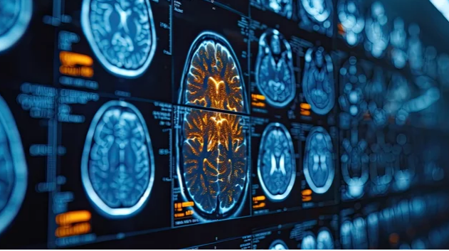
Understanding MRI in Cancer Detection
MRI uses strong magnetic fields and radio waves to create detailed images of internal organs and tissues. Its advantages include:
- High-resolution soft tissue imaging – Essential for detecting tumors in the brain, breast, prostate, and liver.
- Non-invasive nature – No exposure to ionizing radiation.
- Functional imaging – Techniques such as diffusion-weighted imaging (DWI) or dynamic contrast-enhanced MRI (DCE-MRI) provide insights into tissue characteristics.
MRI can detect suspicious areas that may indicate cancer, guide biopsies, and monitor treatment response.
What a Biopsy Does That MRI Cannot
A biopsy involves removing a small tissue sample from a suspicious area for microscopic examination. Biopsies are considered the gold standard for cancer diagnosis because they provide definitive evidence of malignancy.
Key aspects biopsies provide that MRI cannot:
- Cellular and molecular information – MRI shows structure, but only a biopsy confirms whether cells are cancerous.
- Tumor grading and subtype – Histopathology reveals the aggressiveness and type of cancer, guiding treatment.
- Biomarker and genetic testing – Biopsies allow for molecular profiling, important for personalized therapies.
Without tissue confirmation, clinicians cannot be certain of a cancer diagnosis.
When MRI Can Reduce the Need for Biopsies
While MRI cannot fully replace biopsies, it can sometimes reduce unnecessary procedures:
- Prostate Cancer – Multiparametric MRI (mpMRI) can identify lesions that are most likely to be clinically significant. Targeted biopsies can then focus only on these areas, reducing unnecessary sampling.
- Breast Cancer – High-risk women with negative mammograms but suspicious MRI findings may undergo MRI-guided biopsy. In some cases, MRI helps avoid biopsy of clearly benign lesions.
- Liver Lesions – Dynamic contrast-enhanced MRI can help distinguish between benign and malignant liver nodules, sometimes sparing patients from biopsy.
MRI is increasingly used as a triage tool—to determine which lesions truly require biopsy and which can be monitored.
Limitations of MRI as a Replacement for Biopsies
Despite its sophistication, MRI has limitations:
- Cannot confirm malignancy – MRI can indicate suspicious areas but cannot determine cellular characteristics.
- False positives and negatives – MRI may detect benign lesions as suspicious (false positive) or miss small or early-stage tumors (false negative).
- Limited specificity – Some cancers, especially in lungs or gastrointestinal organs, are challenging to identify with MRI alone.
- No molecular or genetic data – Essential for personalized treatment planning.
These limitations make it clear that biopsies remain indispensable for definitive cancer diagnosis.
Emerging Approaches: MRI-Guided Biopsies
One of the most promising strategies combines MRI and biopsy:
- MRI-guided biopsy – Radiologists use MRI to precisely locate suspicious areas, improving the accuracy of tissue sampling.
- Fusion techniques – MRI images are fused with ultrasound or CT during biopsy to target lesions more effectively.
- Reducing unnecessary procedures – Only lesions flagged as suspicious on MRI are biopsied, minimizing patient discomfort and risk.
These approaches maximize the strengths of MRI while retaining the diagnostic certainty provided by biopsy.
Future Perspectives
Advances in imaging and molecular diagnostics may further refine the role of MRI in cancer detection:
- Radiomics and AI – Algorithms can analyze MRI scans for patterns that predict malignancy, potentially reducing reliance on biopsy in select cases.
- Functional MRI techniques – May provide more accurate tumor characterization, helping differentiate benign from malignant tissue.
- Non-invasive liquid biopsies – Blood-based tests combined with MRI may eventually reduce the need for tissue biopsies in specific contexts.
However, even with these innovations, tissue confirmation will likely remain the gold standard for the foreseeable future.
Conclusion: MRI Enhances but Does Not Replace Biopsies
MRI is an extraordinary tool in cancer care, providing detailed imaging that can detect tumors early, guide biopsies, and monitor treatment. However, it cannot replace biopsies because it cannot confirm malignancy, provide tumor grading, or support molecular testing.
The most effective approach integrates both: using MRI to identify and target suspicious lesions while relying on biopsy for definitive diagnosis. This combination improves accuracy, reduces unnecessary procedures, and ensures patients receive the most precise and effective cancer care.
Also Read :