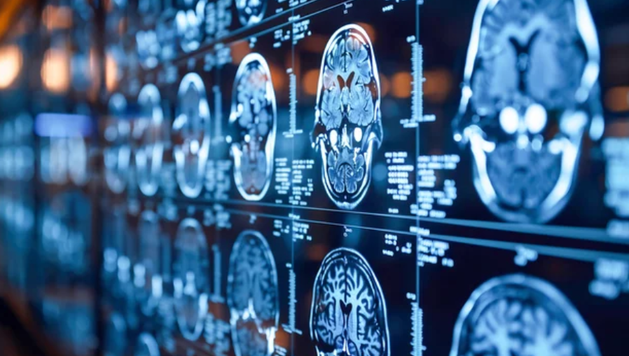
Neurology is entering a new era—one defined by data, precision, and imaging technologies that reveal the brain in extraordinary detail. At the center of this transformation is Magnetic Resonance Imaging (MRI), a tool that has evolved far beyond its original diagnostic purpose. From mapping brain activity to guiding surgical interventions, MRI now stands as a critical pillar of neuroscience and brain health management.
As neurological disorders like Alzheimer’s, Parkinson’s, epilepsy, and multiple sclerosis (MS) continue to rise globally, the need for early detection and personalized treatment is more urgent than ever. MRI’s ability to visualize both structural and functional changes in the brain without radiation exposure makes it the safest and most powerful tool for advancing neurological care.
Looking ahead, the future applications of MRI in neurology promise to reshape how we diagnose, treat, and even prevent brain diseases—turning hospitals into precision-driven centers of neurocare.
The Evolution of MRI in Neurological Care
MRI has long been a cornerstone of neurology, offering high-resolution images of brain anatomy that detect tumors, vascular abnormalities, and degenerative diseases. But in recent years, MRI technology has expanded its capabilities to capture the brain’s dynamic functions—from blood flow and oxygenation to neural connectivity and molecular changes.
Key milestones in MRI evolution include:
- Functional MRI (fMRI): Measures brain activity by detecting blood flow changes, helping scientists map regions responsible for movement, emotion, and cognition.
- Diffusion Tensor Imaging (DTI): Tracks the movement of water molecules in white matter, allowing visualization of brain connectivity networks.
- Magnetic Resonance Spectroscopy (MRS): Detects biochemical changes, offering early insight into neurodegenerative processes.
- High-Field MRI (7T and above): Provides microscopic resolution, revealing subtle lesions or abnormalities invisible in conventional imaging.
These advancements mark a shift from static imaging to dynamic brain analysis, where MRI becomes a window into both structure and function.
Functional MRI: Seeing the Brain Think
Among all MRI innovations, functional MRI (fMRI) has had the most profound impact on neuroscience. By tracking blood-oxygen-level-dependent (BOLD) signals, fMRI allows researchers to see which parts of the brain activate during specific tasks, thoughts, or sensations.
In clinical practice, fMRI is used to:
- Map Brain Function Before Surgery: Neurosurgeons use fMRI to identify critical speech, movement, and sensory areas to avoid during tumor removal or epilepsy surgery.
- Understand Psychiatric Disorders: Studies use fMRI to examine neural networks involved in depression, anxiety, and schizophrenia.
- Monitor Cognitive Decline: fMRI patterns reveal early dysfunctions in Alzheimer’s and other dementias, even before symptoms fully manifest.
In the future, AI-assisted fMRI analysis will enhance our understanding of mental and neurological disorders by decoding complex brain patterns and predicting disease progression.
MRI and Artificial Intelligence: Smarter Brain Diagnostics
The integration of AI and machine learning (ML) into MRI interpretation is ushering in a new era of precision neurology. AI algorithms analyze MRI data faster and more accurately than traditional methods, detecting minute abnormalities that might escape the human eye.
Emerging AI-MRI Applications in Neurology:
- Early Alzheimer’s Detection: AI identifies subtle hippocampal shrinkage and gray matter loss long before clinical symptoms appear.
- Multiple Sclerosis Tracking: Automated lesion segmentation helps neurologists assess disease activity and treatment response.
- Stroke Prediction and Recovery Planning: AI-based MRI analysis predicts stroke risk and guides personalized rehabilitation.
- Brain Tumor Classification: Machine learning differentiates between tumor types, grades, and treatment responses with remarkable precision.
In the near future, AI-powered MRI systems will become decision-making allies—delivering predictive insights, streamlining workflows, and enhancing diagnostic accuracy across every neurological domain.
MRI in Neurodegenerative Disease Management
Neurodegenerative disorders represent one of the greatest challenges in modern medicine. Conditions like Alzheimer’s, Parkinson’s, ALS, and Huntington’s disease often progress silently for years before symptoms emerge. MRI is now being used to detect the earliest structural and functional biomarkers of these diseases, offering hope for earlier intervention.
- Alzheimer’s Disease: MRI tracks hippocampal atrophy and cortical thinning, while advanced MRS detects abnormal chemical accumulation linked to amyloid and tau pathology.
- Parkinson’s Disease: MRI reveals degeneration in the substantia nigra, enabling early-stage diagnosis before severe motor symptoms develop.
- Amyotrophic Lateral Sclerosis (ALS): Diffusion imaging identifies disrupted neural pathways connecting motor regions.
- Huntington’s Disease: MRI monitors progressive gray matter loss and network disconnection across the brain.
With the integration of AI, these MRI biomarkers will soon serve as digital fingerprints of brain health, enabling clinicians to predict, prevent, and slow neurological decline.
MRI-Guided Neurosurgery: Precision in the Operating Room
Intraoperative MRI (iMRI) is one of the most significant technological advances in modern neurosurgery. It allows surgeons to visualize the brain in real time during surgery, ensuring precise removal of tumors or lesions while preserving critical brain functions.
Benefits of MRI-Guided Neurosurgery:
- Real-Time Confirmation: Surgeons can verify that all abnormal tissue has been removed before completing the operation.
- Increased Safety: fMRI maps functional areas, helping avoid damage to speech or motor centers.
- Reduced Repeat Surgeries: Intraoperative imaging minimizes the risk of incomplete resections.
- Enhanced Patient Recovery: Less invasive and more accurate surgeries lead to quicker recoveries and better outcomes.
Future operating rooms will feature AI-assisted MRI navigation systems, providing surgeons with real-time alerts and predictive guidance throughout procedures.
MRI and Mental Health: Visualizing the Invisible
Recent studies have revealed that MRI can detect neurobiological patterns underlying mental health disorders, offering objective data where diagnosis has long relied on subjective assessments.
Functional and structural MRI are being used to study:
- Depression: Altered connectivity between the prefrontal cortex and limbic system.
- Anxiety Disorders: Overactivation of the amygdala and fear-response circuits.
- Schizophrenia: Structural changes in gray matter and disrupted communication between brain regions.
- Autism Spectrum Disorder: Differences in cortical thickness and connectivity affecting social and sensory processing.
The next decade may see MRI integrated into personalized psychiatry, helping tailor treatment strategies based on a patient’s unique neural signature.
Portable and Low-Field MRI: Expanding Global Access
Traditional MRI systems are large, expensive, and confined to major hospitals. However, low-field and portable MRI innovations are making neuroimaging more accessible worldwide.
Compact MRI systems operating at lower magnetic strengths can be installed in smaller clinics or mobile units—bringing advanced brain diagnostics to remote areas and emergency settings.
Future Benefits Include:
- Faster Stroke Diagnosis in ERs: Portable MRI can quickly assess brain damage at the bedside.
- Neonatal and Pediatric Imaging: Safer, quieter scans suitable for infants and children.
- Global Health Access: Low-cost MRI expands access in low-resource regions.
By democratizing neuroimaging, these systems will make brain health monitoring a universal healthcare standard, not a luxury.
The Role of MRI in Brain Research and Innovation
MRI continues to push the frontiers of brain research. Advanced MRI techniques like connectomics, quantitative MRI, and multimodal imaging are helping scientists map how the brain communicates and adapts over time.
The combination of MRI with neuroinformatics and AI modeling enables researchers to simulate brain functions, test treatments virtually, and predict neurological outcomes with unprecedented accuracy.
In the coming years, MRI will play a leading role in developing neuroprosthetics, brain-computer interfaces, and regenerative therapies, bridging the gap between neuroscience and technology.
Challenges and Future Directions
While the future of MRI in neurology is bright, challenges remain:
- High Costs: Advanced MRI systems and maintenance can strain healthcare budgets.
- Complex Data Analysis: Interpreting vast imaging datasets requires AI-driven support.
- Standardization Issues: Varying protocols between institutions can affect data consistency.
However, continuous innovation—especially in AI automation, cloud-based analytics, and quantum MRI—is poised to overcome these limitations, making MRI faster, smarter, and more accessible.
Conclusion: MRI and the Future of Brain Health
MRI is more than a diagnostic tool—it is becoming the central intelligence system for understanding, preserving, and enhancing brain health. From early disease detection to guided neurosurgery and personalized psychiatry, MRI’s role in neurology continues to expand at an extraordinary pace.
The future promises AI-driven, patient-centered neuroimaging ecosystems, where MRI provides not only pictures but also predictive insights into the brain’s structure, function, and future.
As hospitals evolve into precision treatment centers, MRI will remain the gold standard of neurological care—seeing what others cannot, guiding where others hesitate, and unlocking the mysteries of the mind for generations to come.
Also Read :