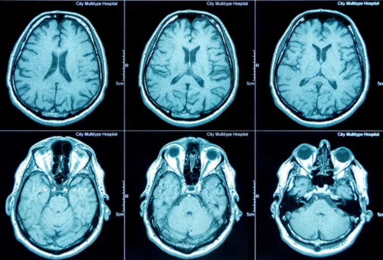
In the world of modern medicine, precision saves lives. Surgeons today demand more than just clear images — they need complete, interactive, and three-dimensional visualizations of the human body. This is where 3D Magnetic Resonance Imaging (3D MRI) is revolutionizing surgical planning. By transforming flat, two-dimensional slices into lifelike anatomical models, 3D MRI empowers surgeons to plan and perform procedures with unprecedented accuracy and confidence.
From neurosurgery to orthopedics, and from oncology to cardiac operations, 3D MRI is reshaping how surgery is planned, practiced, and executed. It is not just a technological innovation — it is a paradigm shift toward safer, smarter, and more personalized medicine.
What Is 3D MRI? The Evolution of Imaging Precision
Traditional MRI scans produce two-dimensional images of internal body structures in multiple slices. While detailed, these images require mental reconstruction by the physician to visualize spatial relationships — a process prone to interpretation errors.
3D MRI, on the other hand, acquires volumetric data from multiple planes simultaneously, allowing computers to reconstruct a detailed, three-dimensional representation of the scanned area. Surgeons can then rotate, zoom, and interact with these 3D models, exploring tissues, vessels, and organs from every possible angle.
This ability to virtually “see” inside the body before surgery transforms how procedures are planned and executed. It bridges the gap between imaging and reality, enhancing surgical safety and precision.
How 3D MRI Is Revolutionizing Surgical Planning
The integration of 3D MRI into preoperative workflows has changed every stage of surgical planning — from diagnosis and simulation to navigation and recovery.
1. Accurate Preoperative Visualization
3D MRI provides a complete spatial understanding of the surgical site. Surgeons can study anatomical relationships in detail — such as how a tumor encroaches on vital blood vessels or how close a lesion lies to a nerve.
This level of visualization allows for precise incision planning, minimal tissue disruption, and optimal surgical approaches tailored to each patient.
2. Virtual Surgery and Simulation
Before making the first incision, surgeons can now perform virtual surgeries using 3D MRI models. These simulations allow them to practice the operation, identify potential challenges, and test different strategies in a risk-free digital environment.
For complex procedures like brain tumor removal or spinal reconstruction, this reduces surprises in the operating room and improves outcomes.
3. Enhanced Navigation During Surgery
Intraoperative guidance is another major breakthrough. By integrating 3D MRI data into surgical navigation systems, surgeons can align real-time visuals with preoperative scans.
This “GPS for surgery” helps track instruments with submillimeter accuracy, ensuring that critical structures are preserved and target areas are precisely reached — even in minimally invasive procedures.
4. Personalized Surgical Strategies
Every patient’s anatomy is unique, and 3D MRI captures these differences with exact detail. Personalized 3D models allow surgeons to customize surgical techniques for each individual — improving fit for implants, optimizing reconstruction geometry, and enhancing functional recovery.
5. Better Communication and Collaboration
3D MRI models are not only useful for surgeons but also for the entire medical team. Radiologists, anesthesiologists, and nurses can share a common visual understanding of the anatomy.
Additionally, 3D images make it easier to explain complex surgical procedures to patients, helping them visualize their condition and understand the planned approach — improving trust and consent.
Applications of 3D MRI Across Medical Specialties
The impact of 3D MRI extends across numerous surgical fields, transforming both planning and execution.
1. Neurosurgery
The brain’s complex structure demands surgical precision at a microscopic level. 3D MRI enables accurate brain mapping, visualizing tumors, blood vessels, and white matter tracts in detail.
Surgeons can simulate craniotomies, plan tumor resections, and avoid critical functional areas, reducing risks of postoperative complications such as paralysis or speech impairment.
2. Orthopedic and Spine Surgery
In orthopedics, 3D MRI provides clear visualization of bones, cartilage, tendons, and ligaments. It aids in joint reconstruction, ligament repair, and spinal fusion planning by offering exact measurements and alignment data.
Custom 3D-printed implants can even be designed directly from MRI models, ensuring a perfect anatomical fit.
3. Cardiac and Vascular Surgery
3D MRI plays a crucial role in cardiac surgery and vascular interventions, where understanding complex vessel networks is essential.
Surgeons use 3D MRI to plan bypasses, repair aneurysms, and guide catheter-based procedures with precision — significantly reducing risks in delicate heart operations.
4. Oncology Surgery
In cancer treatment, 3D MRI enables precise tumor delineation and helps determine surgical margins with millimetric accuracy. This ensures complete removal of malignant tissue while preserving as much healthy tissue as possible — a critical balance for organ preservation and patient quality of life.
5. ENT and Craniofacial Surgery
For head and neck surgeries, 3D MRI helps visualize nerves, muscles, and small anatomical structures in high resolution. Surgeons can plan reconstructive or cosmetic procedures more precisely, achieving superior aesthetic and functional results.
Integration with Augmented and Virtual Reality
The combination of 3D MRI with Augmented Reality (AR) and Virtual Reality (VR) is the next frontier in surgical planning.
By projecting 3D MRI models into AR headsets or VR environments, surgeons can “walk through” the anatomy, exploring every structure in immersive detail.
- AR overlays 3D MRI data directly onto the patient’s body during surgery, guiding incisions and instrument placement.
- VR simulation allows teams to train collaboratively on patient-specific cases before the actual procedure.
These immersive technologies make surgery safer, more efficient, and more interactive — ushering in the era of mixed-reality medicine.
The Benefits of 3D MRI in Surgical Practice
The advantages of 3D MRI extend far beyond visualization. It brings measurable improvements to every aspect of surgical care:
- Enhanced surgical precision and safety
- Reduced operative time through better preoperative planning
- Lower complication rates by minimizing damage to healthy tissues
- Improved postoperative recovery with more targeted interventions
- Better patient communication and education through visual explanations
- Facilitated interdisciplinary collaboration between surgeons, radiologists, and engineers
Ultimately, 3D MRI supports the global shift toward data-driven, patient-specific, and minimally invasive surgery.
Challenges and Limitations
Despite its immense potential, 3D MRI is not without challenges:
- High Costs and Accessibility: Advanced MRI equipment and 3D reconstruction software remain expensive, limiting availability to major hospitals.
- Data Complexity: Processing and interpreting large 3D datasets require powerful computing and specialized expertise.
- Integration Barriers: Combining MRI data with surgical navigation systems and AR platforms requires standardized protocols and training.
However, as technology advances and costs fall, 3D MRI is expected to become mainstream in surgical planning worldwide.
Future Directions: AI and Predictive 3D Modeling
The next evolution of 3D MRI lies in its integration with Artificial Intelligence (AI) and Machine Learning (ML). AI can automate the reconstruction of 3D images, identify surgical landmarks, and even predict postoperative outcomes.
Imagine a future where surgeons receive AI-driven simulations that recommend the safest surgical approach or estimate patient recovery based on 3D MRI data.
These predictive capabilities could redefine how we approach surgery — making it more precise, personalized, and preventive.
Conclusion: A New Dimension in Surgical Precision
3D MRI has changed the way surgeons see and operate. By turning static images into dynamic, interactive models, it enables unmatched precision, confidence, and safety in surgical planning.
From virtual rehearsals to real-time navigation, 3D MRI has become the backbone of next-generation surgery — where data, visualization, and innovation converge to improve lives.
As medicine continues to embrace digital transformation, 3D MRI stands as a cornerstone of surgical evolution, shaping a future where every operation is safer, smarter, and uniquely tailored to the patient.
Also Read :
