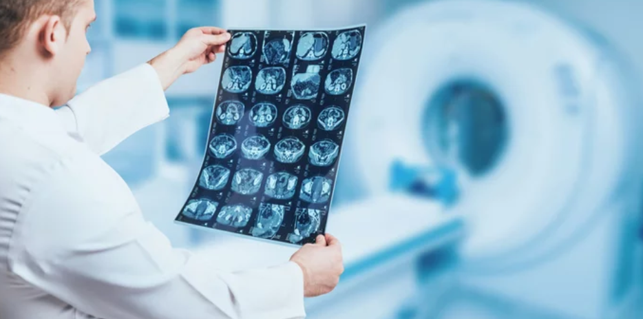
Magnetic Resonance Imaging (MRI) has long been the cornerstone of modern medical diagnostics, offering unparalleled insights into the human body without radiation exposure. Yet, the evolution of MRI technology has reached a groundbreaking new phase — the era of 7 Tesla (7T) MRI. This ultra-high-field imaging system is transforming clinical practice, unlocking details once invisible to even the most advanced 3T scanners. From brain mapping to early disease detection, the 7T MRI stands at the frontier of precision medicine.
What Is 7T MRI? Understanding the Ultra-High Field Power
The term “7T” refers to the magnetic field strength of seven Tesla, which is more than twice that of the widely used 3T MRI systems and nearly 140 times stronger than Earth’s magnetic field. This enormous magnetic strength allows for unprecedented image resolution and contrast, enabling clinicians to observe the human anatomy at a microscopic level.
Unlike conventional MRI systems, the 7T MRI can reveal structural, metabolic, and functional details of tissues that were previously beyond reach. It’s particularly effective in neuroimaging, musculoskeletal studies, and cardiovascular research — areas where visualization of subtle changes can alter clinical decisions.
The Evolution of MRI Technology: From 1.5T to 7T
MRI technology has undergone a remarkable journey since its introduction in the late 20th century. The 1.5T MRI set the standard for clinical imaging for decades, offering reliable anatomical detail. The 3T MRI, introduced later, doubled the magnetic field strength, improving image clarity and reducing scan times.
The leap to 7 Tesla MRI, however, represents a quantum jump in imaging capability. The higher field strength translates to a stronger signal-to-noise ratio (SNR), enabling finer spatial resolution and more accurate tissue characterization. Researchers can now visualize neural networks, microvasculature, and metabolic processes with astonishing clarity — paving the way for earlier and more precise diagnoses.
Clinical Advantages of 7T MRI: Precision Beyond Imagination
The integration of 7T MRI into clinical settings has revolutionized several medical fields. Its enhanced imaging power delivers tangible benefits across multiple specialties.
1. Neurology and Brain Research
7T MRI is a game-changer in neurology, providing ultra-detailed views of the brain’s structure and function. It helps identify small lesions, cortical layers, and microhemorrhages that lower-field systems might miss. Researchers use it to study neurodegenerative diseases such as Alzheimer’s, Parkinson’s, and multiple sclerosis with unmatched precision.
Functional MRI (fMRI) at 7T allows for superior mapping of brain activity, making it invaluable for surgical planning and cognitive research.
2. Musculoskeletal Imaging
In orthopedics and sports medicine, 7T MRI enables visualization of fine cartilage structures, tendons, and ligaments in extraordinary detail. Early signs of tissue degeneration or injury can be detected sooner, improving outcomes for athletes and patients alike.
3. Cardiac and Vascular Imaging
Although challenging due to motion artifacts, advances in cardiovascular 7T MRI have allowed researchers to visualize heart tissue microstructure and vessel walls in ways never before possible. This opens new possibilities for early detection of atherosclerosis, myocardial scarring, and perfusion abnormalities.
4. Oncology and Metabolic Imaging
7T MRI offers significant improvements in tumor detection and characterization. The high-resolution images enable clinicians to distinguish between malignant and benign tissues more accurately. Moreover, its capability for metabolic imaging and spectroscopy supports precision oncology, revealing biochemical changes before visible structural alterations appear.
Technical Innovations Driving 7T MRI Adoption
The transition from research environments to clinical use has been made possible by several key innovations:
- Advanced Radiofrequency (RF) Coils: New coil designs help manage the challenges of ultra-high magnetic fields, improving image uniformity and signal clarity.
- Improved Shimming Techniques: These reduce field inhomogeneities, ensuring better imaging consistency across larger anatomical regions.
- AI-Powered Image Reconstruction: Artificial intelligence now plays a crucial role in optimizing image quality, noise reduction, and diagnostic accuracy.
- Enhanced Safety Protocols: Regulatory advancements and careful engineering have addressed safety concerns, allowing 7T systems to meet FDA and CE clinical standards.
Challenges and Limitations of 7T MRI
Despite its immense promise, the implementation of 7T MRI faces a few hurdles:
- High Cost: The installation and maintenance of a 7T MRI scanner are significantly more expensive than 3T systems, limiting accessibility to large academic and research hospitals.
- Patient Comfort and Safety: Stronger magnetic fields may cause peripheral nerve stimulation or discomfort for some patients. Strict safety guidelines are necessary to ensure patient well-being.
- Technical Complexity: Artifacts and field inhomogeneities remain challenges, particularly for body imaging outside the brain. Continuous improvements in hardware and software are addressing these issues.
However, as technology advances and costs gradually decrease, the mainstream adoption of 7T MRI in clinical practice is becoming increasingly realistic.
Regulatory Milestones and Global Adoption
The U.S. FDA’s approval of the first 7T MRI system (Siemens Magnetom Terra) in 2017 marked a historic milestone. Since then, leading institutions in Europe, Asia, and North America have incorporated 7T MRI into both research and clinical workflows.
Hospitals like the Mayo Clinic, University College London, and Massachusetts General Hospital have reported significant breakthroughs using 7T MRI for neurological and musculoskeletal applications.
As international collaborations grow, 7T MRI is expected to evolve into a clinical gold standard for high-precision imaging.
7T MRI and the Future of Personalized Medicine
The ultra-high resolution and functional capabilities of 7T MRI align perfectly with the goals of personalized and precision medicine. It enables clinicians to:
- Detect diseases at their earliest stages.
- Monitor treatment responses with greater accuracy.
- Tailor therapeutic strategies to individual patient physiology.
In combination with AI analytics, genomics, and molecular biomarkers, 7T MRI represents the next step in integrated, data-driven healthcare.
The Road Ahead: Expanding Clinical Boundaries
The future of 7T MRI looks promising. Ongoing research aims to expand its use beyond brain imaging into whole-body diagnostics, oncology, and cardiology. With the help of machine learning algorithms, image reconstruction speed and accuracy are improving, making scans faster and more patient-friendly.
Moreover, efforts are underway to make 7T MRI more cost-effective and widely available, ensuring that hospitals worldwide can benefit from its diagnostic power.
Conclusion: A New Era in Diagnostic Imaging
The rise of 7T MRI marks one of the most significant milestones in the history of medical imaging. Its unmatched resolution, advanced functionality, and capacity for deep tissue visualization are transforming how clinicians diagnose, understand, and treat disease.
While challenges in cost and accessibility remain, the momentum behind ultra-high-field MRI is unstoppable. As technological innovations continue to mature, 7T MRI will redefine precision medicine, offering clinicians the ability to see — and heal — in ways once thought impossible.
Also Read :
