Medical imaging has revolutionized diagnostics, enabling early detection and precise characterization of various diseases, including cancer. Among the array of imaging modalities, Magnetic Resonance Imaging (MRI) stands out for its detailed soft tissue contrast and lack of ionizing radiation. This article explores the role of MRI in cancer detection, its safety profile, and where it fits within the broader landscape of medical imaging.
Overview of Medical Imaging Modalities
- X-ray and CT (Computed Tomography): Use ionizing radiation; high spatial resolution; effective for bone and lung imaging.
- Ultrasound: Uses sound waves; safe; ideal for soft tissue and fetal imaging.
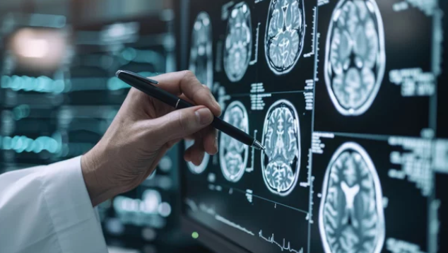
- MRI: Uses magnetic fields and radiofrequency pulses; no ionizing radiation; excellent soft tissue contrast.
- Nuclear Medicine (PET, SPECT): Use radioactive tracers; functional imaging; higher radiation dose.
MRI and Cancer Detection
Advantages of MRI
- High Soft Tissue Contrast: Ideal for brain, spinal cord, liver, and pelvic cancers.
- Multiplanar Imaging: Provides images in multiple planes without repositioning the patient.
- Functional Imaging Capabilities: Including diffusion-weighted imaging (DWI), perfusion, and spectroscopy.
- Lack of Ionizing Radiation: Safer for repeated use, especially in younger patients.
Common Cancers Detected or Monitored by MRI
- Brain and spinal cord tumors
- Breast cancer (especially with dedicated breast MRI)
- Liver tumors (e.g., hepatocellular carcinoma)
- Prostate cancer
- Pelvic cancers (e.g., ovarian, cervical)
MRI and Cancer Risk
Safety Profile
MRI does not involve ionizing radiation, making it inherently safer than CT or nuclear medicine techniques, especially for:
- Repeated imaging: Follow-up assessments
- Pediatric patients: Reduced long-term radiation risk
- Pregnant women: Avoids radiation exposure
Concerns and Limitations
- Contrast Agents: gadolinium-based agents are generally safe, but in rare cases, they can cause nephrogenic systemic fibrosis (NSF) in patients with severe kidney disease.
- Long-term Gadolinium Retention: Emerging research suggests trace amounts of gadolinium may remain in tissues; ongoing studies are assessing clinical significance.
- Cost and Accessibility: MRI is more expensive and less available than other modalities.
Where MRI Fits in with Other Imaging Modalities
- Initial Screening: Often, modalities like mammography or ultrasound are first-line, with MRI reserved for high-risk screening or further characterization.
- Complementary Use: MRI can complement CT or PET scans to provide detailed anatomical and functional information.
- Follow-up and Monitoring: Preferred for assessing treatment response and detecting recurrence, due to lack of radiation.
Future Directions
- Advanced MRI Techniques: Including molecular imaging, hyperpolarized MRI, and artificial intelligence integration.
- Personalized Imaging Protocols: Tailoring scans based on individual risk profiles.
Conclusion
MRI plays a crucial role in the modern diagnostic landscape for cancer detection and management. Its safety profile, especially the absence of ionizing radiation, makes it a valuable tool for initial assessment, detailed tumor characterization, and monitoring. While cost and accessibility remain challenges, ongoing technological advancements promise to expand MRI’s role further, enhancing early detection and improving patient outcomes.
Also Read :