Contribution of fMRI in Neurosurgery: Improvement of Surgical Outcome and Patient Safety
fMRI has transformed neurosurgery by providing highly detailed information on the activity of the brain. It enables neurosurgeons to delineate important areas of the brain for safer and better decision-making during complex surgery. Non-invasive imaging with fMRI has proven indispensable in tumor, epilepsy, and other neurological disorder surgeries.
Due to the fact that neurosurgeons try to minimize risks and maximize patient outcomes, fMRI has become one of the most important parts in pre-surgical planning and intraoperative navigation. By knowing the real-time functioning of the brain, they will be able to avoid critical areas responsible for controlling movement, language, and sensory processing. This novel technique has enhanced surgical success and, simultaneously, ensured better recovery times for patients.
With technological advancement, the application of fMRI is very hopeful in neurosurgery. Further research may mean even better inclusion of fMRI in surgical practice for the care of patients and for better results.
Key Takeaways
- Functional MRI helps in mapping of important regions in the brain before any surgical intervention.
- It maximizes the safety and efficiency of neurosurgical procedures.
- Research is ongoing with efforts to have better technologies for fMRI as related to the improvement of patient outcomes.
Understanding Functional MRI
fMRI is a very powerful tool for neurosurgery and neuroscience. The main purpose of the fMRI is to trace the working areas of the brain and to understand their functions. Some of the main focuses are the technology behind fMRI, mapping of the brain, and how it compares to other imaging techniques.
Principles of fMRI Technology
It works by detecting changes in blood flow and is a reflection of general brain activity; the more active parts of the brain need more oxygen, which requires greater blood flow. fMRI detects this change using a method referred to as BOLD-contrast (Blood Oxygen Level Dependent).
Imaging is done through the use of a fMRI machine while the patient is lying inside a big magnet. This allows a doctor to visualize the activity of the brain without invasion. The images generated show which part of the brain is most active during the execution of different tasks or stimuli.
Brain Mapping and Localization
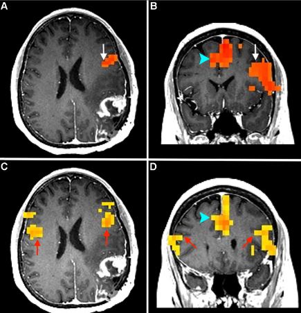
Brain mapping is the process of revealing which part of the brain is responsible for certain functions. fMRI helps in locating the various centers of movement, speech, vision, and many other activities. For example, a doctor can ask a patient to perform some tasks while in the scanning machine so that he can know which section of the brain would be involved in such tasks.
This information is very important to neurosurgeons. It helps them plan surgeries to avoid damaging the necessary areas. A correct localization allows doctors to operate with greater safety and with better results.
fMRI Compared to Other Imaging Modalities
fMRI is an imaging technique quite different from other techniques such as CT and conventional MRI. The CT scan gives exquisite details concerning brain anatomy but does not depict cerebral activity. Standard MRI provides good information about anatomy but lacks the functional aspects.
What is so unique about the fMRI is its ability to monitor the functioning aspect of the brain. It provides a look into the way the brain functions in a dynamic mode, which is quite critical in the development of complex disorders. This very ability guides the medical practitioners in attempting the treatment of disorders such as epilepsy or tumors of the brain.
Functional MRI in Neurosurgical Applications
In neurosurgery, one of the essential tools used is functional MRI. The images help in formulating surgical plans, taking decisions during actual surgery, and evaluating post-surgical outcomes for recovery. This technology helps doctors make out on the working of the brain; therefore making it easier to do the procedures without risk and safely.
Pre-Surgical Planning
fMRI locates vital brain regions and their respective functions during pre-surgical planning. The surgeons may observe sections that are responsible for motion, speech, among other essential tasks. It helps the surgeons not to touch the critical functions while removing a tumor or lesion.
fMRI scans outline the moving part of the brain during specific tasks that a patient will undertake. These results are combined with conventional pictures to give a detailed view of the arrangement of the brain. For example, a surgeon may use data from fMRI to find out how close a tumor is to the motor area.
fMRI does not fail to assist the surgeon in determining a surgical plan tailored to the particular individual. With knowledge of which area of the brain to avoid, various techniques can be selected based on minimizing risks. This therefore leads to better outcomes and less likelihood of complications.
Intraoperative Decision Making
During surgery, fMRI continues to serve an important purpose. Live fMRI monitoring can provide real-time information about brain function. This helps surgeons make immediate decisions if surprises arise during surgery.
For example, in the case of a surgeon mistakenly locating brain tissue near a critical area, fMRI data can inform him/her. They can either proceed with caution or alter the approach they will take. This kind of flexibility may prevent damage to critical functions.
fMRI during surgery helps ensure the brain’s functional integrity is preserved. The surgeons are able to respond appropriately to changes as dictated by the fMRI feedback. This ensures patient safety and provides support for superior surgical outcomes.
Assessment and Monitoring after Surgery
After surgery, fMRI becomes useful in assessing the recovery of the patient. It shows how well the brain has adapted after removing a tumor or lesion. Changes in brain functioning can be monitored by clinicians over time.
The changes can be evaluated by comparing the pre-surgical and post-surgical fMRI scans. This forms vital information in understanding the journey toward the recovery of a patient. It is also of great help in determining strategies for rehabilitation that best fit the needs of the individual.
Moreover, fMRI allows for possible complications to be detected. Early identification of new problems by the medical teams can take timely measures. This can result in quicker recoveries with a better quality of life.

Also Read :
- Advanced MRI Techniques in Neurosurgical Planning
- The Intersection of Biomedical Engineering and Healthcare Innovation
- Tech Innovations in Healthcare: Improving Patient Care with AI
- Medical technology encompasses a wide range of tools, techniques,
- Biomedical Engineering Projects: Innovative Solutions for Healthcare