Magnetic Resonance Spectroscopy: Advances and Applications in Neurosurgical Oncology
Magnetic Resonance Spectroscopy has an ever-increasing important role in neurosurgical oncology. It avails the physician with such precise information at diagnosis and treatment regarding brain tumors that it enhances his decision-making capabilities. MRS goes beyond classical imaging into the measurement of the chemical composition of tissues, offering additional information on the type of tumor and its nature.
Clinicians, surgeons, and oncologists use MRS for differential diagnosis of tumors prior to surgical intervention, and follow-up studies to evaluate the response of treatment. This type of modality will go a long way towards enhancing patient outcomes, making care personalized. Specific biochemical markers of tumors form the very basis for targeted therapies so that better survival rates could be ensured.
MRS technology is continually evolving, and this would more than likely enhance its contribution to neurosurgery even further. This will not only help diagnosis but will also allow surgical intervention and postoperative management to become much more effective.
Take Home Points
- MRS offers details on the chemical composition of brain tumors.
- The neurosurgical patients have a better diagnosis along with treatment planning
- There is improvement in patient outcomes with the development of MRS technology
Principles of Magnetic Resonance Spectroscopy
The contribution of MRS to the analysis of metabolic changes in the brain is similarly an advanced imaging technique. Basic principles guide this technology, just like in MRI. Main issues to take into account include the physical principles of the technology, technological advances, and how to interpret the metabolite peaks.
Physical Principles of MRS
Magnetic Resonance Spectroscopy uses powerful magnets and radio waves to study the chemical composition of tissues. Within such a magnetic field, protons within tissues start vibrating at peculiar frequencies.
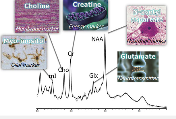
These frequencies depend on what type of chemical environment each proton finds itself in. On the addition of a radio frequency pulse, MRS detects the signals emitted from the very start. This gives insight into the various metabolites present in the brain, such as choline, creatine, and N-acetylaspartate.
These frequency variations create a spectrum. Each metabolite will appear as a peak at a different location in this spectrum. The area underneath each peak corresponds to the concentration of that metabolite, and thus enables one to perform quantitative analysis.
Technological Advance of MRS Techniques
There have been considerable technological advances in MRS techniques over the years. One important innovation has been higher strength-of-field magnets. These increase SNR so that smaller concentrations of metabolites are easier to detect.
Multivoxel spectroscopy allows the measurement of spatial resolutions, thus enabling various regions of the brain to be studied simultaneously.
Software improvements and advances in data processing also enhance MRS interpretability. Methods such as automatic frequency correction serve to refine the spectra into clearer, more distinct readings.
Interpretation of Metabolite Peaks in MRS
Peak interpretation of the metabolites is an essential part of MRS analysis. Each metabolite appears at a specific chemical shift given in parts per million (ppm). Some of the important metabolites include:
- Cho (choline): This is a marker of cellular membrane turnover. It is generally increased in tumors.
- Cr: Creatine serves as the reference. It indicates energy metabolism.
- NAA: This is a marker for neuronal health. Low levels could indicate neuronal loss.
These peaks, in turn, provide very important patterns and ratios. For example, an increase in the choline to NAA ratio may indicate increased tumor activity. Preciseness of interpretation assists the clinician in diagnosis and treatment response monitoring in patients with brain tumors.
Applications of MRS in Neurosurgical Oncology
MRS thus plays an important role in the management of brain tumors, since the application enhances decision-making on surgery and improves outcomes in patients. The outcome of the techniques allows the clinician to make better assessments of the tumor at various stages of treatment.
Preoperative Planning and Tumor Grading
MRS offers metabolic information in brain tumors that are helpful during preoperative planning. Differentiating types of tumors such as gliomas and metastases is done by certain metabolites. The main metabolites include choline, creatine, and N-acetylaspartate.
High levels of choline usually accompany heightened activity of the tumor, and low levels of N-acetylaspartate are indicative of neuronal loss. This analysis of metabolites will assist the surgeon in assessing the grade of the tumor thus making informed decisions on treatment. Tailor-made strategies can be planned by the surgical teams once such reports are received and it leads to enhanced accuracy of surgery.
Intraoperative Evaluation and Guidance
MRS may help the surgical team intraoperatively during surgery. It has information on tumor margins and details of critical brain structures. These are important to avoid inflicting injury to the brain while performing tumor removal.
Surgeons can use MRS to assess whether the tumor has been satisfactorily resected completely. The technique allows distinguishing between residual tumor tissue and normal brain tissue. Immediate feedback provided by MRS creates an improvement in the outcomes of complex surgical procedures.
Postoperative Assessment and Treatment Monitoring
After surgery, MRS assists in evaluating the treatment outcome. It allows monitoring metabolic alteration of the brain that contributes to the early diagnosis of recurrence of tumor. Different scans reveal metabolic markers that signal the activity of the tumor.
MRS can also guide further treatment decisions, such as the need for radiation or chemotherapy. This modality offers continuous information to the healthcare team in order to make prudent decisions on the care of the patients. These often result in timely interventions that improve not only survival but quality of life.

Also Read :