Advances in Diagnosis and Care
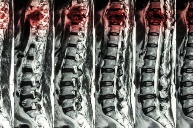
MRI for Spinal Cord Injury and Neurosurgical Treatment, by R. Ramakrishna et al. MRI has become an indispensable tool in the diagnosis of spinal cord injury and neurosurgical treatment. This imaging modality allows the treating physician to visualize the spinal cord at high resolution and make appropriate diagnoses and surgical plans. Understanding how MRI functions in this setting may significantly enhance the capability of the patient and caregivers to make informed decisions regarding their care.
Spinal injuries tend to have quite devastating effects on a person’s life and therefore need prompt treatment. A clear view from the injury MRI scan will help the doctor in deciding on an appropriate method of treatment. The right information will ultimately allow the patient to make some informed decisions regarding their alternative options that are available for treatment and the expected outcomes.
Neurosurgical interventions may follow the MRI evaluation in the treatment of spinal cord injury. These procedures can help pain, mobility, and overall function. Knowing how MRI informs these interventions will help you participate in your recovery actively.
Take Home Message
- MRI plays an important role in the diagnosis of spinal cord injury.
- Sharp images enable appropriate neurosurgical interventions.
- Knowledge of MRI facilitates patient decision-making.
Principles of MRI for Spinal Cord Injury
MRI plays an indispensable role in the diagnosis of spinal cord injury cases. It helps in the identification of the extent of the damage, thereby guiding the treatment options.
Overview of MRI Technology
Magnetic Resonance Imaging, commonly called MRI, is a non-invasive method of taking pictures of the body using strong magnets and radio waves. It does not involve the use of X-rays or any other forms of radiation, hence making it very safe for use on many patients.
This process entails lying inside a big, tunnel-like machine. The machine aligns the water molecules in the body and produces highly detailed pictures of the spinal cord and tissues around it. MRI can visualize soft tissues, which is very important when assessing for nerve damage and inflammation.
The standard protocols of spinal imaging in general include several sequences in order to provide multiple views and types of tissue contrast.
Benefits of MRI in Spinal Diagnostics
Therefore, MRI has a number of advantages in the diagnosis of spinal cord traumas: high-resolution pictures with information about the anatomy of the spinal cord, nerve roots, and surrounding structures are obtained.
This is highly useful for the establishment of soft tissue traumas that other diagnostic techniques may not be able to identify and thus provide substantial information about the nature of injury.
MRI also helps in studying and monitoring changes over a given period. It will be able to show the progress a condition might take and therefore aid in treatment planning. Another advantage is that it is noninvasive; patients will, therefore, be comfortable during their exams.
Since MRI does not use radiation, it is safer for repeated use, especially on young patients.
Challenges and Limitations
Despite its usefulness in many ways, MRI does have some challenges. For example, it can be very noisy inside the machine, and claustrophobic patients will have a problem being inside the tunnel.
MRI is not possible for all patients due to implants or even pacemakers since it interferes with the magnetic field.
Another is the time taken to perform the scan; it takes a maximum of 30 minutes, which may be unsuitable for all patients.
Finally, diagnosis from MRI depends on the proficiency of the radiologist who is examining the image. Hence, this produces discrepancies in diagnosis.
Neurosurgical Treatment Following MRI
After the MRI, the doctors do the planning for surgery based on the results. It includes analysis of the extent of the injury, choosing the appropriate techniques, and planning the postoperative care.
MRI Result Explanation for Surgery
MRI reveals the most essential information about the spinal cord and the tissues around it. The presence of any compression, swelling, or abnormalities is considered by the physicians.
The main points they concentrate on include:
Location of Injury: Knowing which portion of the spinal cord is injured.
Extent of Injury: It gives an idea about the extent of injury so that a clear surgical approach can be planned.
Teamwork: Neurologists and surgeons have to combine their efforts for proper interpretation of results.
These variables will help surgeons make informed decisions concerning surgery.
Surgical Spinal Repair Techniques
After having clear MRI results, surgeons take up appropriate techniques for repair. Few standard techniques include:
Laminectomy: The removal of part of the vertebra to relieve the spinal cord of pressure.
Discectomy: Herniated disc material is removed that may impinge on the spinal nerves. Spinal Fusion: Two or more vertebrae are joined to provide stability to the spine. Techniques are selected by the surgeon based on the type of injury presented and specific patient needs. Each of these methods has an objective of alleviating pain and facilitating healing.
Postoperative Care and Monitoring
Surgery requires monitoring postoperation as recuperation depends on it. Steps followed by doctors and nurses are excluded in ensuring recovery.
Pain Management: Pain is managed through medication. Mobility Assistance: Early physical therapy is administered to gain momentum in the patient’s mobility.
Follow-up appointments provide an avenue through which doctors can observe follow-through in healing and make necessary adjustments to care. Monitoring includes watching for complications such as infection or embolism. This post-operative care will go a long way in making the road to recovery as smooth as possible.
Also Read :
- Magnetic Resonance Spectroscopy in Neurosurgical Oncology
- Functional Connectivity Mapping in Preoperative Neurosurgical Planning
- The Impact of Diffusion Tensor Imaging on Neurosurgical Procedures
- Neurosurgical Tumor Resection and MRI Imaging
- Intraoperative MRI : Revolutionizing Neurosurgical Precision