Advanced MRI Sequences in Neurosurgical Interventions: A Step Toward Precision and Outcomes Advanced MRI sequences are yet another important tool in modern neurosurgical interventions. These techniques enhance image quality and provide critical information guiding surgical decisions with regard to improving patient outcomes. Understanding how advanced methods work can substantially help surgeons and other medical professionals better recognize and deal with complicated cases in the brain.
Special MRI sequences will allow physicians to better visualize the tumor, blood vessels, and brain tissue. This will facilitate better surgical planning and execution. As neurosurgery continues to evolve, improvements in imaging will have a much greater impact on enhancing safety and effectiveness.
Advanced MRI sequences are a symbol of not only the development of technology but also a new epoch of more accurate and personalized surgical treatment. It is impossible to stay aside and not be informed about these changes for anyone who takes part in neurosurgery.
Key Takeaways
- Advanced MRI sequences enhance neurosurgical imaging
- Better clarity allows for superior surgical planning.
- Newer techniques provide safer surgical outcomes for the patients.
Bases of Advanced MRI Sequences
Advanced MRI sequences are increasingly assuming prominence in neurosurgical interventions, given their value in providing detailed visualization that helps with decisions for diagnosis and treatment. Knowledge of the evolution, principles, and main sequences of the imaging modality serves to permit the effective harnessing of these techniques.
Principles of MRI Technology
MRI scans utilize powerful magnets and radio waves to generate images of the body. Inside the MRI scanner, hydrogen atoms in the body align with the magnetic field. Then, radiofrequency pulses temporarily knock them out of kilter. As the atoms realign, they emit signals, which are recorded and used to create images of the body.
By using MRI, different tissues are sensitive to this magnetic field and radio waves; therefore, high-resolution anatomic images of the brain and other structures can be developed. Tumors are usually bright compared to normal tissue and thus more easily detectable. The fact that settings can be changed-be it magnetic strength or time of signaling-allows customization based on clinical needs.
Evolution of MRI in Neurosurgery
Neurosurgical applications of MRI have evolved quite a bit since its invention. Early generations of MRI machines were relatively rudimentary in their capabilities and resolution. Advances in technology soon incorporated high-field strength magnets into everyday machines, along with improved coils. This evolution allowed for increased image clarity, as well as reduced scan times.
The addition of functional MRI enabled surgeons to visualize the activity of the brain. This is particularly important in tumor-removing surgeries, as it avoids any damage to the vital portion of the brain. Diffusion tensor imaging enhanced knowledge about white matter tracts. It offers surgeons the opportunity to plan operations for illnesses such as epilepsy or traumatic brain injuries.
Commonly Used MRI Sequences in Neuroimaging
A number of such specific MRI sequences are crucial for neuroimaging. First, T1-weighted images provide a good overview of the anatomy and can thus be used to assess brain structures. Accordingly, the areas containing fluid and pathology, such as oedema or lesions, become prominent in T2-weighted images.
Fluid-attenuated inversion recovery sequences suppress the signal of cerebrospinal fluid in order for the lesions abutting fluid-containing structures to be better delineated. Changes in water diffusion-an indicator of acute ischemia or stroke-are shown by diffusion-weighted imaging.
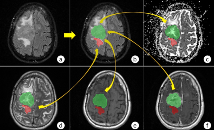
Contrast-enhanced imaging, with the injection of a contrast agent, has improved tumor and vessel delineation. Each one of these sequences provides different information that leads to neurosurgical decision-making and improves outcomes.
Also Read :