
fMRI and Brain Mapping: How Neurosurgical Decision-Making is Improved
fMRI has changed the way physicians think about the brain as to its functionality. It provides crucial information that aids in making neurosurgical decisions by explaining the location of areas in the brain that are important for executing a particular higher order task. The technology guarantees safety and efficacy in surgical procedures of the brain for improved patient outcomes.
What surgeons basically need to know while preparing for such complex procedures is which parts of the brain control important functions, such as movement and speech. fMRI allows them to observe such regions in action in real time, thus enabling the exact planning of surgical intervention. This strategy minimizes the risk of damaging areas that have important roles in the recovery and daily life of a patient.
The role of fMRI in brain mapping is ever-increasing, opening new frontiers in innovative neurosurgical operations. By incorporating recent technology into practice, healthcare professionals can make informed choices to ensure safety and well-being for the patients.
Key Takeaways
fMRI has been found instrumental in the mapping of brain functions related to vital areas to be operated on.
This technology helps in accurately planning neurosurgery, ensuring more safety for the patients.
It results in improved outcomes from neurosurgical procedures due to its application.
Principles of Functional MRI
fMRI is a powerful technique to investigate activity in the brain. It enables researchers and clinicians to determine which brain region is involved in any given function. This chapter discusses the principles underlying the technology of fMRI, neurovascular coupling, and, lastly, problems regarding spatial and temporal resolution.
Principles of the fMRI Technology
Functional MRI relies on changes in blood flow to detect brain activity. A region of the active brain will consume more oxygen than an inactive one. Changes in blood oxygenation will be detected by the MRI using a process called the Blood Oxygen Level Dependent, or BOLD, contrast.
fMRI makes use of strong magnetic fields and radio pulses to produce the best images of the brain. Images of the brain are generated by modifying the signal emitted from hydrogen atoms in the water within the brain. This can be performed completely non-invasively, with the added benefit of not exposing a person to radiation.
Neurovascular Coupling and BOLD Contrast
Neurovascular coupling means the interaction between activity in the brain and blood flow. The action of neurons results in signals that contribute to the dilation of blood vessels in the vicinity. This dilatation increases blood flow into the active area, bringing more oxygen.
The BOLD signal detects the property of oxygenated and deoxygenated hemoglobin that is paramagnetic. The more active areas will have higher signals because more oxygen will be supplied to those areas. This relationship between activity and oxygenation helps the scientist understand which parts of the brain are involved in specific tasks.
Resolution in Space and Time
The spatial resolution refers to the precision with which an fMRI scan can determine where in the brain activity is taking place. Modern fMRI technology typically has a spatial resolution of about 2-3 mm. Thus, one can generally observe the activation of large brain regions, but not the fine details of the same region.
Temporal resolution is the speed at which changes in brain activity can be captured; fMRI has a temporal resolution of roughly 1-2 seconds. This is considered sufficient for most applications; however, changes in activity sometimes can occur in periods less than this.
This modality is used, among others, for planning neurosurgical procedures. Functional MRI identifies critical brain areas involved in motor functions, language, and sensory processing. Information provided by this imaging modality enhances surgical success and greater safety for the patient.
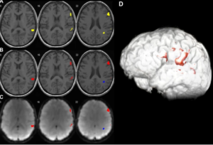
Brain Mapping
Preoperative brain mapping uses functional MRI to find the critical areas of the brain. Surgeons will look at scans that identify specific parts of the brain associated with speech, movement, and sensation. This is done as a means of formulating a personalized surgical plan aimed at preventing critical injury to important parts of the brain.
Some of the specific techniques include:
- Activation Mapping: This outlines regions that are activated in a particular activity.
- Connectivity Analysis: This will present the communicative ability between different parts of the brain.
Both techniques inform the surgeon how to stay away from critical areas so as to maximize the chance of a good outcome.
Assessment of Neuroplasticity
The fMRI helps assess neuroplasticity-or the brain’s ability for reorganization and adaptation. This becomes crucial in patients suffering from brain tumors or lesions. Once the extent of accommodation that has taken place in the brain is known, a physician can plan an approach.
For example, if there is considerable neuroplasticity in a patient, then surgeons will consider using less invasive methods. They will try to save as much healthy brain tissue as possible without compromising on the solution.
Neuroplasticity knowledge helps in recovery forecasts, too. This helps the expert to show patients what to expect during the rehabilitation course realistically.
Analyzing Risks and Benefits
Before surgery, a proper analysis of risks and benefits is necessary. Functional MRI is used to gain substantial information about the functioning of the brain that will help in decision-making.
Physicians balance the benefit of tumor resection against possible risk. Possible risk could be impairment in function due to surgery or from the surgery itself.
- With information gained by using functional MRI, the surgical team evaluates the following:
- Possible complications: In what area of the brain is there a risk to language or movement?
- Predicted benefits: How will quality of life improve if the tumor is removed?
This helps the patient understand what the results of the treatment may be and makes informed choices about care.
Also Read :
- 16. “Functional MRI and Brain Mapping in Neurosurgical Decision-Making”
- Advanced MRI Sequences in Neurosurgical Interventions
- MRI for Spinal Cord Injury and Neurosurgical Treatment
- Magnetic Resonance Spectroscopy in Neurosurgical Oncology
- Functional Connectivity Mapping in Preoperative Neurosurgical Planning