Imaging Biomarkers in MRI for Neurosurgical Outcomes: Improvement in Predictive Accuracy and Treatment Strategies
Imaging biomarkers in MRI are of critical importance in the light of neurosurgical outcomes. These imaging biomarkers assist physicians in the forecast and attainment of better results after brain surgery. Through MRI image analysis, a health team is able to capture key features of the brain which may essentially affect the outcome.
Imaging biomarkers become increasingly more sophisticated as technology evolves. In turn, decision-making during surgical planning and the management of the patient becomes ever more knowledgeable. Understanding these tools greatly impacts how patients will recover and will live beyond neurosurgery.
This blog post discusses the pivotal role imaging biomarkers will have and their relation to successful surgical outcomes. Insights from MRI scans allow for a deeper understanding of the conditions of the brain and therefore enable better treatment for the patients.
- Imaging biomarkers make it possible to predict surgical outcomes.
- Advances in MRI technology translate to better patient care and recovery.
- Biomarkers can aid in fine-tuning surgical planning and decisions.
The Basics of Imaging Biomarker in MRI
Imaging biomarkers represent an important features in MRI for assessing conditions of the brain and predicting the outcome after surgery. They enable the understanding of the anatomy and physiology of the brain and, therefore, are key information for neurosurgeons.
Imaging Biomarkers: Definition
Medical imaging biomarkers are a set of features that are measurable from medical images. They describe the disease or treatment effects in the brain, including specific patterns of brain lesions or regional volumetric changes in the brain. These biomarkers are identified and quantified from MRI studies to provide insight into the patient condition.
These biomarkers help the clinician in differentiating various disorders of the brain. This helps a lot in decision-making and allows personalized treatment. The temporal evaluation of such changes is very useful to assess the efficacy of treatment.
Role of MRI in Neurosurgery
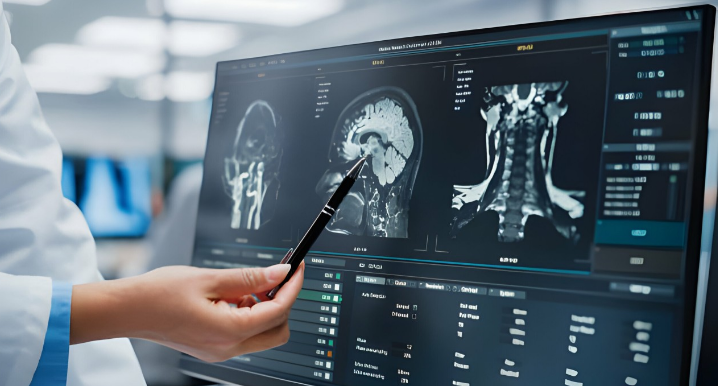
In the near future, MRI will become very important in neurosurgery in terms of procedural planning and guidance. It produces detailed pictures of the brain, which are necessary to outline tumors, lesions, or other abnormalities. High-resolution images give surgeons the exact locations to minimize risks during surgery.
MRI technologies, like fMRI, enable the neurosurgeon to map the activity of a patient’s brain. This is particularly significant for those surgeries in which activity might be occurring near areas of the brain concerned with speech or motor functions. MRI also assists in post-surgical care; through it, surgeons may detect post-operative complications or review the status of healing.
Advances in MRI Technology
Recent advances in MRI technology have now made it possible to attain a high degree of precision for biomarkers using imaging. High-field MRI and DTI are some of the newer techniques that help improve the clarity of the brain scans. These new developments allow for much better visualization of changes in microstructure and connectivity in the brain.
The MRI data is also analyzed with the help of machine learning tools more effectively than before. They can identify pattern recognition in ways that might be difficult or even impossible to do with the naked eye. Advanced imaging combined with technology enhances predictive capability regarding surgical outcome and thereby offers better patient care.
Neurosurgical Outcome Assessment
Neurosurgical outcome assessment must consider several factors. These include the success of resecting the tumor, prognosis for patients, and monitoring of the disease.
Assessment of Tumor Resection Success
Success in the resection of tumors is of essence to ensure good patient outcomes. MRI biomarkers help surgeons in assessing if the tumor is being taken out whole.
The commonly used techniques are DWI and contrast-enhanced MRI. These tools outline regions where tumor cells could still persist.
A clear postsurgical scan indicates a good resection. Residual tumor may imply complications or the recurrence of tumor growth. Continued follow-up via MRIs keeps surgeons ready to act quickly, if needed.
Predicting Patient Prognosis
Patient prognosis is largely based on the findings made during preoperative MRI. The size, location, and type of tumor become really significant.
Imaging biomarkers may reveal how aggressive a tumor is likely to be. For instance, higher rates of certain biomarkers can indicate a poor prognosis.
Being able to identify these risks helps health professionals to plan treatment options. Knowledge of the potential risks concerning various tumor types can facilitate tailored patient care.
Monitoring Disease Progression
Monitoring of disease progression is another role of post-surgery MRI. Continued imaging allows the physicians to witness any changes, if at all, in the patient’s condition.
A good example could be the rise in tumor markers due to recurrence.
This helps in the quantification of these changes by use of MRI analysis. Further management decisions can be made by the doctors based on this data.
This is quite vital in follow-up, which improves the long-term outcomes for patients.

Also Read :
- MRI in Hydrocephalus and Neurosurgical Shunt Placement
- Intraoperative MRI and the Future of Neurosurgical Navigation
- Functional MRI and Brain Mapping in Neurosurgical Decision-Making
- MRI for Spinal Cord Injury and Neurosurgical Treatment
- The Impact of Diffusion Tensor Imaging on Neurosurgical Procedures