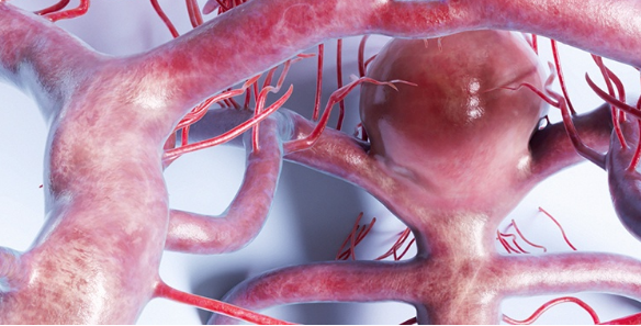
MRI of Cerebral Aneurysms and Neurosurgical Clipping: Techniques and Outcomes Cerebral aneurysms are serious and have the potential to be life-threatening. Accurate detection and management of cerebral aneurysms are crucial for the improvement in outcomes among patients. Cerebral aneurysms are significantly detected by MRI and provide guidance to neurosurgical clipping.
The use of MRI therefore allows great detailing in the imaging of the brain, hence helping the doctors to know the size and location of an aneurysm. This is important information needed when one is deciding on treatment options. Neurosurgeons can then use the results from MRI in accomplishing the clipping safely and effectively.
With enhancement in MRI techniques, the future of dealing with cerebral aneurysms appears even brighter, with clearer images and better decision-making for healthcare professionals.
Key Takeaways
- MRI is indispensable in the precise diagnosis of cerebral aneurysms
- The modality offers enough details for safer neurosurgical clipping procedures
- Cerebral aneurysm treatment is improved with advanced techniques in MRI
Detection of Cerebral Aneurysms: the Role of MRI Technology
The technology behind MRI plays a crucial role in the diagnosis of cerebral aneurysms. MRI produces sharp images of the brain’s structural anatomy without exposing the patient to radiation. Due to the advancement of methods used in MRI, detection and diagnosis have become significantly improved, thus making this imaging modality invaluable in neurosurgery.
Improvement of MRI Techniques
Recent development within the MRI techniques has quite substantially enhanced the diagnosis of cerebral aneurysms. With the advent of the 7.0 Tesla, MRI allows for the delivery of high-resolution images, no doubt enhancing the clear visibility of even minute brain structures.
Other techniques like time-of-flight magnetic resonance imaging and magnetic resonance angiography with enhancement by contrast provide a clear display of the blood vessels; thus, allowing for the identification of even smaller aneurysms that might otherwise remain hidden from conventional methods of imaging.
These developments not only help in diagnosis but also assist in pre-surgical planning. Neurosurgeons can more effectively determine the size and location of aneurysms, which enhances interventions.
MRI Compared to Other Imaging Techniques
MRI has unique merits in comparison with other imaging methods in the diagnosis of cerebral aneurysms. MRI does not make use of ionized radiation, unlike computed tomography. This is much more significant for those patients who require frequent imaging due to the potentially associated long-term hazards.
MRI also holds superior soft tissue contrast. This feature enables clear visualization of the details in structures of the brain and nearby vessels, hence making determination of aneurysms easier. While CT angiography is often quick, MRI remains considered the gold standard in detailed analyses.
It is in these situations that MRI is particularly useful when other imaging tests may be limited. It is sensitive to slight changes in the cerebral vessels, which could have been caused by the presence of an aneurysm.
Role of MRI in the Diagnosis of Aneurysm
MRI thus finds its place in diagnosing cerebral aneurysms. It identifies the aneurysm as well as its characteristics. Details include size, shape, location, and others that are necessary for treatment decisions.
The ability to visualize the surrounding brain tissue also helps identify related complications like hemorrhage. This wide view then helps the physician evaluate the urgency of treatment.
In addition, MRI can be used for follow-up evaluations after treatment. It is important in the management of patients to monitor the stability of an aneurysm or follow-up for new formations. MRI will provide additional information to help further management.
MRI-Guided Neurosurgical Clipping
MRI furnishes an invaluable modality in the management of cerebral aneurysms at the time of clipping. Indeed, it is greatly helpful preoperatively in planning, intraoperatively as guidance, and postoperatively in the assessment of surgical outcome.
Preoperative Evaluation by MRI
Complex MRI scans before the surgery are used to outline the size, shape, and location of an aneurysm. This is necessary to help the surgical team develop an individualized approach in treatment.
This modality also delineates the surrounding brain structures and vessels, hence enabling more informed risk assessment. The kind of images produced-for instance, 3D reconstructions-provides surgeons with an enhanced understanding of the nature of the aneurysm and its relationship to vital brain areas, thereby enhancing precision during the surgical approach.
Intraoperative Use of MRI
Intraoperative MRI does have some growing applications in neurosurgery. This allows the surgeon, while operating, to view real-time images to make changes immediately if needed. This even enables confirmation of whether complete clipping of the aneurysm has been achieved and what changes are occurring during the surgical operation.
This can also reduce the additional need for other surgeries through the use of intraoperative imaging. Moreover, it increases assurance about the outcome, as surgeons can confirm their moves inside the operating theater.
Follow-up and Postoperative Evaluation: MRI after surgery forms the basis of assessing the success of clipping. It needs to be followed by follow-up scans to make sure that the aneurysm remains secure without complication such as rebleeding or changes in the surrounding tissues.
They will often set up a schedule for regular MRI follow-up studies after surgery to ensure that any new developments are caught in their early stages. This is a necessary precaution for the patient’s safety and long-term recovery. Quite often, patients will have a schedule of follow-ups based on individual risk factors and specifics related to their aneurysm.
Also Read :