MRI of Traumatic Brain Injuries: A Neurosurgical Approach Traumatic brain injuries are the most frequent type of injury, having quite devastating health consequences. It is a big challenge for the neurosurgeon to correctly assess such injuries to be able to manage the case appropriately. MRI therefore happens to be quite handy in the assessment of traumatic brain injuries, since it provides quite detailed imagery helpful in detailing the extent of the damage.
It is important to understand how MRI enhances assessment in TBIs for effective treatment. Advanced imaging offers a lead on surgical decisions and rehabilitation that may affect patient outcomes. With clinical applications, professionals can enhance their efforts towards the improvement of outcomes in patients with brain injuries.
Key Takeaways
- MRI gives, in detail, important imaging when it comes to traumatic brain injury assessment.
- Accurate assessment has paved the way for the enhancement of treatment strategies to improve patient outcomes.
- Understanding the application of MRI aids neurosurgeons in making informed decisions.
MRI Technology in Traumatic Brain Injury
MRI technology has, over time, played a critical role in assessing traumatic brain injuries. The technology provides detailed images of the brain structure, enabling neurosurgeons to understand fully the severity and type of injury.
Principles of MRI
MRI and other such imaging are done by using strong magnets combined with radio waves to produce pictures of the brain. It does this by sensing the changing alignment of hydrogen atoms, which are present in all body tissues. Once these atoms are put under a magnetic field, they align uniformly due to that field. A radio frequency pulse collapses this alignment, and while returning to their usual alignment, the atoms emit signals.
These signals are acquired in order to produce images. MRI is particularly good at demonstrating soft tissue and, thus, it is a good modality for diagnosing brain injuries like contusions, hemorrhages, and edema. MRI does not use ionizing radiation; therefore, it is a safer modality if repeated imaging is needed.
Improvements in MRI
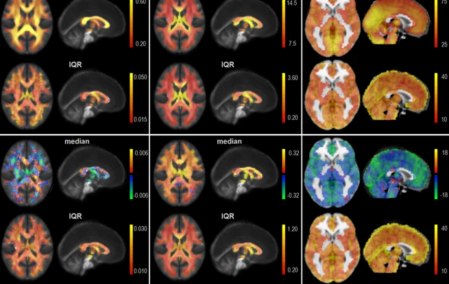
Advanced MRI techniques have significantly improved the diagnosis of brain injuries. An example includes DTI, which can detect the motion of water in tissue within the brains. It can find tiny changes at the level of the microstructure that might not be visible with conventional MRI studies.
fMRI detects changes in blood flow to provide information about brain activity and, thus, how an injury may affect the functioning of the brain. High-resolution MRI techniques also permit more detailed images to characterize the injury.
These have helped in advancing the level of diagnosis and treatment planning. The future of MRI in traumatic brain injury will be an expanding role as technology continues to unfold.
Clinical Applications of MRI
MRI has become a crucial modality in assessing traumatic brain injuries by neurosurgeons. It visualizes brain structures, identifies tissue damage, and is also employed in monitoring recovery.
Assessment of Brain Structures
MRI provides high-resolution detail to the appearance of brain structures, which helps the neurosurgeon in assessing the extent of an injury. High-resolution images can make neurosurgeons detect those areas that have been affected in the brain by trauma.
Key features reviewed include:
- Cerebral edema: There may be swelling that may indicate the severity of the injury.
- Hemorrhage: There may be bleeding, which might necessitate immediate attention.
- Infarction of the brain: An area of dead tissue due to the absence of blood supply.
- By observing these features, surgeons can design specific treatment approaches .
Tissue Injury Detection
MRI offers the all-important detection of subtle and overt tissue injury. Some techniques, such as diffusion-weighted imaging delineate areas of restricted motion of water, thus demonstrating injuries that are not obvious.
The kind of injury to be identified includes:
- Axonal injury: The injury of nerve fibers that can lead to chronic problems.
- Contusions:Bruises of the brain that may affect function.
- Microbleeds: Tiny leaks that may be missed with other imaging.
Early identification of such injuries assists in making appropriate and timely decisions for intervention.
Recovery and Rehabilitation Monitoring
MRI plays an important role in monitoring the recovery of patients with traumatic brain injuries. Periodic scanning gives some insight into the healing process and the success of rehabilitation processes.
These are followed by key aspects, such as:
- Resolution of edema, which generally indicates progress with recovery.
- Changes in lesion size: This helps to evaluate the effectiveness of the performed treatment.
- Neuroplastic changes: It helps in identifying the way the brain adapts over time.
These insights shall be useful to health care providers, who at all times can adjust these treatment plans based on individual progress and needs.

Also Read :
