
Functional MRI in Neurosurgical Research: Progress and Applications
Functional MRI is one of the strongest tools for neuroscience in peering into the activity of the brain. It visualizes changes in blood flow and thus helps researchers understand how different parts of the brain cooperate. In neurosurgical research, fMRI will help in locating important brain functions before surgery to avoid risks and improve patient outcomes.
In addition to mapping those parts of the brain involved in motion and speech, researchers also use fMRI to detect abnormal activities of the brain associated with pathologies such as tumors and epilepsy. This imaging modality is important in assessing the safety of patients and in the formulation of appropriate treatment strategies. This technique is non-invasive; hence, it offers a number of advantages compared to the older techniques, and this makes the technique very vital in the modern study of neurosurgery.
With the continuous improvement in the technique of fMRI, the list of its applications is also growing. Neurosurgeons and researchers continue to explore new ways of utilizing this modality to further understand the brain and enlighten neurosurgical practice. Combinations of fMRI and neurosurgery will transform patient care and treatment.
Key Takeaways
- fMRI stands out as a very crucial part in understanding activity and function of the brain.
- It maps essential areas of the brain prior to surgery and increases the safety of the patient.
- Continual developments enhance its applications in neurosurgical practices.
Fundamentals of Functional MRI
Functional MRI is a brain research and surgical tool in neuroscience. It serves to visualize the activity of the brain and to assist decisions in clinical situations. This section addresses the core principles of the technology behind fMRI, its use in preoperative planning, and the role that it plays in mapping the brain to understand its function.
Principles of Technology
fMRI works by detecting changes in blood flow in the brain. This technique relies on the blood-oxygen-level dependent (BOLD) contrast, which measures the level of oxygen in the blood. Whenever there is activity in a certain area of the brain, more oxygen is required; hence, the flow of blood increases.
The scanner uses powerful magnets to generate images. These images show the different areas of the brain and their relative activity when different functions are performed. The information received can be used to build a picture of how various areas of the brain work together.
Key properties of fMRI include:
- Non-invasive-surgery or needles are not involved.
- Real-time imaging-it shows the activity of the brain as it is happening.
- High spatial resolution-it can pinpoint activity in specific parts of the brain.
fMRI in Preoperative Planning
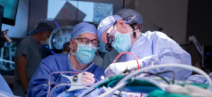
fMRI has an important role in neurosurgical planning. Before surgery, the surgeons identify, with the use of fMRI data, those parts of the brain which are responsible for the most important functions. In this way, they try not to cause any damage to those parts of the brain during the operation.
fMRI scans can map language, movement, and sensation. By knowing the location of these functions, surgeons may make more informed decisions. This approach enhances the possibility of a successful surgery with less post-surgical side effects.
fMRI in preoperative planning is done by:
- Locating functional areas: The maps indicate the important regions.
- Surgical decision-making: Information guides about where to operate.
- Minimizing risks: avoids compromising significant functions of the brain.
Mapping of the Functional Brain
Functional brain mapping is an imperative approach towards the understanding of the interactions of various parts of the brain. fMRI has enabled the study of these interactions visually while an individual is performing some task or even at rest. This type of mapping is an important aspect of research on disorders of the brain, cognitive processes, and normal brain function.
These maps help to identify different networks involved in various tasks, including:
- Memory
- Emotion
- Movement
Such maps are also of immense benefit in designing therapies that could reach specific parts of your brain. It helps us understand things about the function of your brain that regular imaging couldn’t tell us, which improves your outcome.
Also Read :