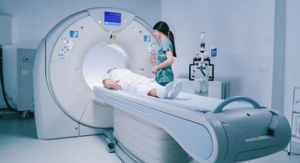
MRI scans allow doctors to see inside the body without surgery by creating detailed images of tissues and organs. This technology captures data beyond what the human eye can detect and uses advanced visual tools to turn that data into clear, meaningful pictures. The power of MRI lies in its ability to reveal hidden structures through sophisticated imaging techniques that make the invisible visible.
Recent advances in visual technology have improved how MRI images are displayed and understood. Innovations like 3D models, augmented reality, and enhanced visualization methods help doctors and researchers experience MRI data in new ways, making it easier to diagnose and treat diseases. These tools bring deeper clarity to complex medical images, transforming raw data into interactive visual stories.
By enhancing the ways MRI data is viewed, medical professionals gain better insights that can lead to faster decisions and more accurate treatments. This blend of imaging and visual tech is opening new doors for medical science and patient care, making invisible health issues clearly visible.
Key Takeways
- MRI reveals internal body structures without surgery through detailed imaging.
- Visual tools help transform MRI data into clear, interactive images.
- Advanced imaging tech supports better diagnosis and treatment decisions.
MRI Technology and Visual Innovation
MRI uses powerful magnets and radio waves to create detailed images of the body’s interior. Over time, the way these images are produced and processed has improved greatly. This has helped medical professionals see smaller details and better understand the human body.
Principles of Magnetic Resonance Imaging
MRI works by aligning hydrogen atoms in the body using a strong magnetic field. When radio waves pulse through, these atoms emit signals. The MRI machine detects these signals and converts them into images.
The key elements are the magnetic field strength, radiofrequency pulses, and signal detection. Stronger magnets allow clearer images. MRI does not use harmful radiation, making it safer than some other imaging methods.
This method can show soft tissues very clearly, which helps with diagnosing conditions in organs, muscles, and the brain. The images are created in slices, which can be combined to form 3D views.
Evolution of MRI Visualization
Early MRI machines produced basic black and white images with limited detail. Over the decades, improvements in magnet strength and scanner design have made images sharper and faster to capture.
Newer machines use larger bores, making scans more comfortable. Visual quality has also improved by increasing the magnetic field strength and adding advanced coils.
The shift from simple contrast to detailed, dynamic imaging means doctors can now observe tissue function and blood flow in real-time. This evolution has made MRI a vital tool in both diagnosis and research.
Advances in Image Processing
Modern MRI uses advanced software to enhance images. Algorithms improve clarity and reduce noise, helping doctors spot abnormalities earlier and more accurately.
AI plays a growing role, analyzing images to highlight areas of concern and assist in making treatment decisions. Image reconstruction techniques have also become faster, allowing quicker results.
Additional features like color mapping and 3D visualization help medical professionals better understand complex structures. These advances improve both diagnosis and patient outcomes by providing clearer, more detailed views.
Visual Tech Enhancing MRI Experiences
Advancements in visual technology have changed how MRI scans are done and understood. They improve image clarity, offer new ways to view data, and help patients feel calmer during scans. These tools combine software and hardware to create more interactive, detailed, and understandable MRI experiences.
3D Imaging and Interactive Displays
3D imaging takes MRI data beyond flat images. It builds detailed, three-dimensional models of the scanned area, which doctors can rotate and explore from different angles. This helps in better understanding complex structures, such as organs or tumors, assisting in diagnosis and treatment planning.
Interactive displays often use touchscreens or special software. They allow medical staff to zoom in, highlight areas, and compare images side-by-side. This hands-on experience makes it easier to spot changes over time or plan surgeries.
These 3D models can also be shared with patients. Showing clear visuals helps patients understand their condition and the steps needed for care. It bridges the gap between technical medical data and patient knowledge.
AI-Powered Image Interpretation
Artificial intelligence (AI) is now used to analyze MRI scans quickly and more accurately. AI software can detect patterns or abnormalities that might be missed by the human eye. It helps radiologists prioritize urgent cases by flagging critical areas.
AI also supports consistent image reading, reducing errors caused by fatigue or inexperience. It can enhance image quality by reducing noise and improving contrast, which leads to clearer pictures.
Additionally, AI helps predict possible health outcomes based on imaging data. This kind of support assists doctors in making timely and informed decisions, improving patient care.
Augmented and Virtual Reality Applications
Augmented reality (AR) and virtual reality (VR) transform how people interact with MRI data. AR overlays MRI images onto the patient’s body or surgical field, providing real-time guidance during procedures.
VR offers full immersion by simulating the MRI environment or visualizing internal body structures in 3D. It also serves as a tool to reduce patient anxiety. Patients can experience a virtual tour of the MRI process or view calming scenes while being scanned.
Both AR and VR systems help medical teams plan surgeries with greater precision and educate patients more effectively. These technologies turn complex MRI information into clear, interactive experiences that support better outcomes.
Also Read :
