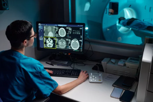
The integration of Augmented Reality (AR) with Magnetic Resonance Imaging (MRI) is transforming the way surgeons plan, navigate, and perform complex procedures. By combining high-resolution MRI scans with immersive AR overlays, doctors can visualize a patient’s anatomy in real time—enhancing precision, reducing risk, and improving surgical outcomes.
This powerful combination is ushering in a new era of image-guided surgery, where the surgeon’s view of the operating field is enriched with accurate, interactive 3D anatomical maps. In this article, we’ll explore how AR and MRI work together, their applications in modern surgery, and what the future holds for this groundbreaking medical innovation.
The Need for Enhanced Surgical Visualization
Surgery has always relied on accurate anatomical knowledge. However, traditional imaging methods—such as 2D MRI scans or CT images—require surgeons to mentally translate static images into the three-dimensional reality of a patient’s body. This cognitive step can be challenging, especially for intricate procedures involving the brain, spine, or cardiovascular system.
AR and MRI integration addresses this challenge by:
- Providing real-time 3D anatomical overlays on the surgical field.
- Allowing surgeons to see inside tissues and structures without making large incisions.
- Enabling precise navigation to avoid damaging critical structures.
How AR and MRI Work Together
- Preoperative MRI Scanning
High-resolution MRI scans are taken before surgery to capture detailed anatomical data, including soft tissue, blood vessels, and structural abnormalities. - 3D Reconstruction
Specialized software converts MRI data into interactive 3D models. These models can be color-coded to highlight key areas—such as tumors, nerves, or blood vessels. - Augmented Reality Integration
Using AR devices (such as Microsoft HoloLens or AR-compatible surgical microscopes), the 3D MRI data is overlaid directly onto the patient’s body during surgery. - Real-Time Surgical Guidance
Surgeons can manipulate the overlay—rotating, zooming, or adjusting transparency—to gain deeper insight while maintaining focus on the operative field.
Applications in Modern Surgery
1. Neurosurgery
AR-MRI integration is especially valuable in brain surgery, where precision is critical. Surgeons can:
- Navigate complex brain structures.
- Avoid damaging functional areas like motor and speech regions.
- Plan minimally invasive paths to deep-seated tumors.
2. Orthopedic Surgery
In joint replacement and spinal surgeries, AR overlays of MRI scans help align implants accurately and avoid nerve injury.
3. Cardiac Surgery
MRI visualizations combined with AR guide surgeons through delicate heart procedures, ensuring safe access to areas that are otherwise difficult to view.
4. Oncology Surgery
In tumor removal, AR-enhanced MRI helps identify the exact boundaries of cancerous tissue, increasing the chances of complete removal while sparing healthy structures.
Benefits of AR-MRI Surgical Visualization
- Increased Accuracy – Precise anatomical mapping reduces surgical errors.
- Shorter Surgery Times – Real-time guidance streamlines decision-making.
- Less Invasive Procedures – Smaller incisions and targeted interventions reduce recovery time.
- Better Training Tools – Medical students and residents can learn through immersive, real-world simulations.
Challenges and Limitations
While the AR-MRI combination is promising, some challenges remain:
- High Cost – Advanced AR systems and MRI equipment require significant investment.
- Technical Complexity – Integrating imaging data into AR platforms demands specialized software and calibration.
- Learning Curve – Surgeons need training to fully leverage AR’s capabilities.
The Future of AR-MRI in Surgery
Emerging trends suggest even more advanced possibilities:
- Real-Time Intraoperative MRI – Scanning during surgery to update AR overlays instantly.
- AI-Powered Visualization – Machine learning algorithms to automatically highlight surgical targets.
- Remote AR Surgery – Combining AR and telemedicine for expert-guided procedures across the globe.
With continuous improvements in imaging resolution, computing power, and AR hardware, the fusion of MRI and augmented reality is set to become a standard feature in operating rooms of the future.
Conclusion
The marriage of Augmented Reality and MRI represents a monumental leap forward in surgical visualization. By providing surgeons with a detailed, interactive view of a patient’s anatomy in real time, this technology not only improves precision but also enhances patient safety and recovery.
As adoption grows and technology advances, AR-MRI integration will redefine surgical standards—turning complex operations into safer, faster, and more efficient procedures. The future of surgery is not just in the surgeon’s hands—it’s also in the clarity of their vision.
If you’d like, I can create an SEO-optimized meta description and keyword set for this AR-MRI surgical visualization article so it ranks well in search engines. Would you like me to prepare that next?
Also Read :
