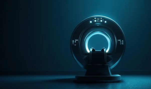
In the ever-evolving world of medical education, interactive MRI simulators are emerging as one of the most transformative tools for teaching anatomy, physiology, and diagnostic skills. By combining high-resolution magnetic resonance imaging with advanced visualization technology, these simulators offer a dynamic, hands-on learning experience that traditional textbooks and static images simply cannot match.
For students, MRI simulators bridge the gap between theoretical learning and real-world clinical practice, providing a safe environment to explore anatomy, interpret images, and understand disease processes—without the pressure of working on live patients.
Why Interactive MRI Simulators Are Changing the Game
Traditional MRI training often begins during clinical rotations, leaving students with limited exposure before entering real-life patient care. Interactive MRI simulators address this gap by allowing students to:
- Explore anatomy in multiple planes (axial, sagittal, coronal).
- Manipulate and rotate 3D reconstructions for a better spatial understanding.
- Practice MRI interpretation in a self-paced, risk-free setting.
- Simulate pathology progression to learn how diseases evolve visually.
This hands-on approach doesn’t just teach students what to look for—it trains them how to think like a clinician.
Core Features of Modern MRI Simulators
1. Realistic MRI Data Sets
Simulators use authentic MRI scans from anonymized patient cases, giving students exposure to real-world variations in anatomy and pathology.
2. 3D Visualization Tools
Interactive platforms allow learners to slice, rotate, and zoom into high-resolution anatomical structures, making it easier to understand complex spatial relationships.
3. Guided Learning Modules
Many simulators integrate built-in tutorials and quizzes, helping students check their progress and strengthen weak areas.
4. Pathology Libraries
These collections showcase a variety of medical conditions—such as tumors, strokes, and joint injuries—allowing learners to compare normal and abnormal anatomy side-by-side.
5. Integration with VR and AR
Virtual reality (VR) immerses learners in a life-sized anatomical space, while augmented reality (AR) overlays MRI scans onto physical models for hybrid learning experiences.
Benefits for Medical Students and Educators
For Students
- Active Learning: Hands-on interaction improves memory retention compared to passive reading.
- Diagnostic Skills: Early exposure to MRI interpretation builds confidence for future clinical roles.
- Adaptability: Students can learn at their own pace, revisiting scans as needed.
For Educators
- Efficient Teaching: Simulators allow instructors to present complex cases without needing live patients.
- Standardized Learning: Every student gets access to the same high-quality MRI datasets.
- Performance Tracking: Built-in analytics help educators measure student progress.
Applications Beyond the Classroom
While MRI simulators are primarily used in medical schools, they also serve a purpose in:
- Radiology residency training for advanced diagnostic skill-building.
- Surgical planning practice to visualize anatomy before entering the operating room.
- Continuing medical education for practicing physicians.
- Patient education, helping individuals better understand their conditions.
Challenges and Considerations
Despite their benefits, MRI simulators require careful planning for successful integration:
- High Initial Costs: Quality simulators and 3D visualization systems can be expensive.
- Technical Training: Students and faculty need orientation to use advanced tools effectively.
- Data Security: Patient scans must be anonymized to protect privacy.
The Future of MRI Simulation in Education
The next generation of MRI simulators will likely include:
- AI-driven interpretation guides that highlight key areas of interest on scans.
- Global collaborative learning platforms where students can analyze the same case in real time across different countries.
- Gamification features that turn diagnostic learning into interactive challenges.
With these advancements, MRI simulators will not only enhance learning but also prepare medical students to excel in a technology-driven healthcare landscape.
Conclusion
Interactive MRI simulators represent a revolution in medical education, offering students an engaging, immersive way to learn anatomy, understand pathology, and develop diagnostic expertise. By merging visual technology with real-world clinical data, they create a bridge between classroom learning and patient care—shaping a generation of healthcare professionals who are both technically skilled and visually literate.
In the coming years, as technology becomes more affordable and widespread, interactive MRI simulators are poised to become a standard in medical training worldwide.
Also Read :
