Radiology is rapidly evolving, driven by technological advancements that are reshaping how medical imaging is interpreted, taught, and applied. Among the most promising innovations are the integration of magnetic resonance imaging (MRI) and augmented reality (AR) in radiologist training. This combination is transforming the way future specialists learn to analyze complex imaging data—making education more immersive, accurate, and efficient than ever before.
MRI provides unparalleled detail in imaging soft tissues, while AR overlays this data into an interactive, 3D space. Together, they give radiologists-in-training the ability to visualize anatomy, pathology, and spatial relationships in ways that static 2D images cannot.
Why Radiologist Training Needs Innovation
Traditional radiology education relies heavily on 2D images displayed on screens, lectures, and static textbooks. While effective to a degree, this approach often limits:
- Spatial understanding of anatomical structures.
- Hands-on experience with complex and rare pathologies.
- Real-time collaboration between instructors and learners.
Given the increasing complexity of imaging studies and the demand for precise interpretations, training methods must evolve to equip radiologists with the skills needed in a data-heavy, high-tech healthcare environment.
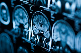
The Role of MRI in Modern Training
MRI remains one of the most versatile imaging modalities, capable of providing:
- High-resolution, multi-plane images for detailed analysis.
- Functional imaging capabilities (such as fMRI) to study brain activity.
- Contrast-enhanced imaging for better visualization of tumors and lesions.
For radiologists in training, MRI datasets offer a realistic foundation to learn about anatomy and pathology in exceptional detail.
How Augmented Reality Transforms MRI Learning
AR technology enhances MRI-based training by overlaying digital content into the real world through headsets or smart displays. Here’s how AR adds value:
1. 3D Anatomical Exploration
Students can view MRI scans reconstructed into 3D models, rotate them, and explore internal structures layer by layer.
2. Interactive Pathology Visualization
AR allows trainees to compare healthy anatomy with diseased states in a side-by-side, immersive view.
3. Real-Time Collaboration
Instructors and students can interact with the same AR-enhanced MRI model from different locations, enabling remote yet engaging training.
4. Procedure Planning Practice
Trainees can simulate interventions such as biopsies or surgical approaches using AR overlays of MRI data.
Benefits for Radiology Education
Enhanced Spatial Understanding
3D visualization helps radiologists grasp complex anatomical relationships more intuitively.
Higher Engagement and Retention
AR transforms passive learning into active exploration, leading to better knowledge retention.
Exposure to Rare Cases
Trainees can interact with MRI scans of uncommon pathologies that they might never encounter in real life.
Safe, Controlled Learning Environment
Mistakes made in AR simulations carry no risk to patients, allowing learners to practice freely.
Applications Beyond Training
While its primary role is in education, the combination of MRI and AR is also being applied in:
- Surgical planning for neurosurgery, orthopedics, and oncology.
- Patient education, allowing individuals to see their own scans in an understandable 3D format.
- Collaborative diagnostics, where teams of specialists work together in virtual environments.
Challenges to Implementation
Despite its promise, integrating MRI and AR into radiologist training has obstacles:
- High equipment costs for advanced AR headsets and visualization software.
- Technical learning curves for both students and faculty.
- Data privacy concerns with patient scans.
- Infrastructure requirements for handling large MRI datasets.
The Future of MRI and AR in Radiology Training
As AR hardware becomes more affordable and MRI datasets more accessible, we can expect:
- AI-assisted MRI interpretation within AR platforms to guide learners.
- Cloud-based learning hubs where radiologists worldwide can share and analyze cases in real time.
- Haptic feedback integration, allowing users to “feel” tissue resistance in simulated procedures.
The synergy between MRI and AR is not just an educational tool—it’s a vision of the future of medical imaging.
Conclusion
Training radiologists with MRI and AR technology is revolutionizing medical imaging education. By combining the precision of MRI with the immersive interactivity of AR, educators can offer richer, more engaging learning experiences that prepare radiologists for the complexities of modern healthcare.
As the technology becomes more accessible, it is poised to become a standard component of radiology training programs worldwide—bridging the gap between theoretical learning and real-world expertise.
Also Read :