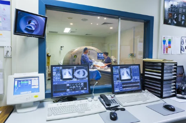
Magnetic Resonance Imaging (MRI) has long been a cornerstone of modern medical diagnostics, offering unmatched clarity and detail in visualizing the human body. Traditionally, MRI scans were confined to radiologists’ screens, with complex grayscale images interpreted by specialists. Today, visual learning tools are transforming how these scans are displayed, understood, and applied—not just for medical professionals, but also for students, patients, and researchers.
The journey from scan to screen now involves advanced visualization software, 3D reconstructions, augmented reality (AR), and even virtual reality (VR). This evolution is making MRI data more interactive, intuitive, and educational than ever before.
The Shift Toward Visual Learning in Medicine
Historically, MRI results were delivered as static slices—two-dimensional cross-sections that required years of training to interpret accurately. While effective for seasoned radiologists, this format posed challenges for:
- Medical students trying to grasp complex anatomical relationships.
- Patients attempting to understand their own conditions.
- Specialists collaborating across different fields and locations.
By adopting visual learning techniques, MRI data can now be transformed into accessible, interactive content that bridges the knowledge gap between experts and learners.
From Raw Scan to Interactive Display
The modern MRI workflow now incorporates advanced stages of visual processing:
1. Image Acquisition
MRI scanners capture detailed images of soft tissues, organs, and even functional processes such as brain activity.
2. Post-Processing
Specialized software reconstructs raw MRI data into 3D models, enhancing clarity and spatial understanding.
3. Interactive Visualization
These models can be displayed on high-resolution monitors, AR headsets, or VR platforms, enabling real-time exploration.
Key Visual Learning Tools for MRI Data
3D Anatomical Reconstructions
Instead of flipping through dozens of 2D slices, learners can manipulate lifelike anatomical models, zoom into specific regions, and view structures from multiple angles.
Augmented Reality Overlays
AR technology projects MRI data into real-world space, allowing clinicians or students to view internal anatomy superimposed on a physical mannequin or patient.
Virtual Reality Simulations
VR immerses learners in a fully digital environment where they can “walk through” the brain, heart, or musculoskeletal system.
Interactive Reporting Systems
Modern MRI reports include annotated images, color-coded overlays, and clickable elements that explain findings in plain language.
Benefits of Visual Learning with MRI
1. Improved Spatial Understanding
3D and AR tools help learners visualize complex anatomical relationships more intuitively.
2. Better Patient Communication
Visual MRI reports allow patients to see and understand their own scans, increasing trust and compliance with treatment plans.
3. Enhanced Retention for Students
Interactive tools turn passive observation into active exploration, improving memory retention and clinical reasoning skills.
4. Collaborative Learning
Cloud-based MRI platforms enable multiple users to examine the same dataset in real time, regardless of location.
Applications Across Medical Fields
Visual learning with MRI extends beyond radiology:
- Surgery – Surgeons use preoperative 3D MRI models to plan complex procedures.
- Neurology – Brain mapping from MRI scans helps identify functional areas before operations.
- Orthopedics – 3D joint reconstructions aid in implant design and surgical navigation.
- Oncology – Tumor visualization in 3D assists in targeted therapy planning.
Challenges in Scan-to-Screen Transformation
While the benefits are clear, adopting these tools isn’t without hurdles:
- High equipment and software costs.
- Data storage and processing demands for high-resolution MRI datasets.
- Training requirements for both educators and clinicians.
- Privacy and compliance concerns when sharing patient scans.
The Future of MRI-Based Visual Learning
Emerging technologies promise to push MRI visualization even further:
- AI-driven segmentation to automatically highlight key anatomical structures.
- Real-time MRI visualization during surgeries.
- Haptic feedback integration to simulate the tactile feel of tissue in virtual environments.
- Metaverse-style collaborative spaces where medical teams can meet inside a shared 3D MRI environment.
Conclusion
Visual learning with MRI represents a significant leap in how medical imaging is interpreted, taught, and communicated. By moving from scan to screen—and now to interactive, immersive platforms—MRI is no longer just a diagnostic tool; it’s a dynamic educational and communication medium.
As technology continues to evolve, these advancements will make MRI data more accessible and engaging, empowering healthcare professionals, students, and patients alike with a deeper understanding of the human body.
Also Read :
