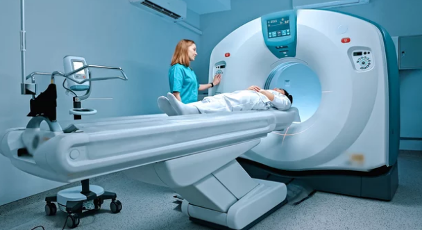
Magnetic Resonance Imaging (MRI) changed medical care. It shows soft tissues like nothing else. It uses no radiation. But MRI creates a lot of complex data. This data needs smart ways to be read. Visual computing is changing MRI research. It blends computer graphics, computer vision, and image processing. This mix pushes limits in understanding our bodies. It helps us see diseases better. This article explores how visual computing opens new doors. It goes from better image creation to smart analysis.
MRI data is huge and complicated. Old methods often miss small, important details. Visual computing helps fix these problems. It uses advanced math and computer power. Researchers are finding new ways to make images clear. They are also making scans faster. This helps us learn more about biology.
We will look at how visual computing changes MRI research. It impacts image reconstruction. It helps with dividing parts of an image. It aids in lining up images. It also helps make new ways to view data. These steps lead to better diagnoses. They help create personal treatment plans. They also improve our grasp of brain issues, cancer, and heart health.
Advancements in MRI Image Reconstruction
AI-Powered Reconstruction Algorithms
Artificial intelligence (AI) helps make MRI images. Machine learning, especially deep learning, turns raw data into clear pictures. This process happens even with less data. What are the benefits? Scans become much faster. The images also get a better signal. This means less noise. So, doctors get clearer pictures faster. This saves time for patients and hospitals.
- Deep Learning Architectures for MRI
Different AI programs help with MRI reconstruction. Convolutional Neural Networks, or CNNs, are great for images. Recurrent Neural Networks, RNNs, handle sequences of data. Generative Adversarial Networks, GANs, can create realistic images. These tools learn from many past scans. They get better at spotting patterns. This makes new MRI images look sharp. They can even fill in missing details.
- Compressed Sensing MRI
Compressed sensing is a smart math trick. It lets us capture less raw data. Yet, it still makes a high-quality image. Visual computing algorithms make this happen. They reconstruct images from fewer measurements. This cuts down the scan time a lot. Imagine an MRI that takes half the time. This is a big win for busy hospitals. It also means less time for patients in the scanner.
Super-Resolution Techniques
Visual computing can make MRI scans look even sharper. This is called super-resolution. It helps us see tiny details. It can reveal very small problems. This means doctors might find issues sooner. It helps spot little lesions.
- Learning-Based Super-Resolution
Deep learning models are now used for MRI super-resolution. These models learn how to add detail. They do much better than old ways of making images clearer. They can turn a blurry scan into a sharp one. This means more useful images from existing scanners. They help doctors see things that were once hidden.
Enhanced Image Analysis and Segmentation
Automated Segmentation of Anatomical Structures
Visual computing helps computers find body parts. It uses methods like atlas-based segmentation. Machine learning and deep learning also play a big role. These tools automatically outline organs, tissues, or problems. This saves doctors a lot of time. It also makes results more reliable every time. This helps avoid human error.
- Deep Learning for Segmentation Accuracy
CNNs, like the U-Net model, are very good at this. They can accurately draw lines around brain parts. They also segment tumors or heart chambers. These models learn from many examples. They get very precise. Many studies show they beat human efforts. This helps in mapping out disease spread.
- Multi-Modal Segmentation
Visual computing can combine different MRI scans. For example, it might use T1-weighted and T2-weighted images. It also uses diffusion-weighted scans. Sometimes, it adds other image types. This mix of information improves how well we segment. It is super helpful for tough cases. This helps doctors get a full picture.
Quantitative MRI and Radiomics
Visual computing lets us get numbers from MRI scans. These numbers show things like water movement or blood flow. They also describe tissue types. This is called quantitative MRI. Radiomics is a newer field. It pulls many different details from medical images fast. It helps us see features a human eye might miss.
- Feature Extraction and Machine Learning
Algorithms can find textures, shapes, and brightness patterns. These come from the segmented areas. Machine learning then uses these details. It can tell the difference between healthy and sick tissue. It predicts how a disease might act. It also helps guess how well a treatment will work.
- Applications in Oncology
Radiomics in MRI is changing cancer care. It helps describe tumors. It can predict how a patient will respond to chemotherapy. It also estimates risks of cancer coming back. This helps doctors pick the best treatment for each person. It makes cancer care more personal.
Advanced Visualization and Interaction
3D Reconstruction and Rendering
Visual computing builds 3D models from 2D MRI slices. These models show body parts and diseases in full 3D. Special rendering techniques make them look real. They make anatomy clearer. They also highlight problems. This gives doctors a better view of complex areas.
- Real-time Volumetric Rendering
It is hard to show huge MRI data sets in 3D right away. But computers are getting faster. New methods allow real-time 3D views. Doctors can spin and slice the 3D images. This lets them look at anatomy from any angle. It makes exploring complex cases easier.
- Augmented Reality (AR) and Virtual Reality (VR) in MRI
AR and VR are big new tools. Surgeons can use AR during operations. It overlays MRI data right onto the patient. VR creates full virtual worlds. Doctors can practice surgeries in these worlds. Patients can use VR to understand their condition better. This helps everyone prepare for what’s next.
Interactive Exploration and Data Fusion
New software lets doctors play with MRI data. They can move it around. They can blend different types of MRI scans. They can even add other patient info. This helps them find new insights. It makes complex data easy to handle.
- Visually Guided Data Exploration
Pictures and interactive tools guide users through data. They help spot small patterns. They show how different things connect. This can lead to new discoveries. It makes research more efficient.
- Integration with Electronic Health Records (EHRs)
Imagine seeing MRI findings right next to a patient’s history. Visual computing makes this possible. It connects images with other health records. This gives a complete view of the patient. It helps doctors make very specific treatment plans. This is key for personal medicine.
Image Registration and Tracking
Non-rigid Registration for Longitudinal Studies
Doctors need to compare MRI scans over time. This helps track diseases. It shows how treatments work. Non-rigid registration handles changes in the body’s shape. It can line up scans even if organs move or change. This is key for long-term health studies.
- Deformable Image Registration Algorithms
Algorithms like B-spline or fluid registration help line up images. They use math to adjust for body changes. They are great for brain scans over many years. They also track heart motion. These tools ensure that we are comparing apples to apples.
- Validation and Accuracy Metrics
It’s important to know how well these tools work. There are ways to check their accuracy. Special numbers tell us how much error there is. This ensures doctors can trust the results. It makes research findings more solid.
Motion Correction and Artifact Reduction
Patients sometimes move during an MRI. This can blur the images. Visual computing helps fix these blurry scans. It uses image registration and other clever methods. These methods clean up the pictures after the scan.
- Real-time Motion Tracking and Correction
New systems can watch patient motion as it happens. They fix the image right away. Or they adjust the scan during the process. This means fewer bad scans. It makes the MRI process smoother. It leads to better images the first time.
Future Directions and Challenges
Integration of Multi-Modal Imaging and Non-Imaging Data
What if we could combine MRI with CT scans or PET scans? Visual computing can blend all these images. It can also add genetic data. It can even use a patient’s health history. This gives a full view of a disease. It helps us see the big picture.
- Data Harmonization and Visualization
Bringing together different kinds of data is tough. But visual computing can make it work. It helps us see how diverse data connects. This can reveal complex links in biology. It helps researchers find answers faster.
Real-time Feedback and Adaptive Imaging
Imagine an MRI machine that learns as it scans. It could change settings on the fly. It could make the image better as it takes it. Or it could focus on specific body events. This is called adaptive MRI. It makes scans smarter.
- Closed-Loop Imaging Systems
These are very smart imaging systems. They learn from the pictures they take. They use this knowledge to scan better next time. This makes MRI faster and more useful. It means more clear diagnoses for patients.
Ethical Considerations and Data Privacy
Using AI in medicine brings up important questions. Patient data must stay private. We need rules and checks for AI tools. These tools must be proven safe and effective. It’s about building trust in new tech.
Conclusion: The Visual Future of MRI
Visual computing is much more than showing MRI pictures. It’s now a key part of MRI research. It drives new ideas from start to finish. It speeds up scanning. It makes images clearer. It helps with deep analysis. It also allows for interactive views. These methods are changing how we use MRI data. AI, smart math, and strong visualization tools are coming together. This promises an MRI future with deeper insights. It means more personal care for each person. This leads to earlier diagnoses and better treatments. Ultimately, it brings better results for patients. The new frontiers are here, and they look amazing.
Also Read :
