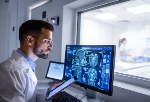
Modern medicine faces a big problem. Doctors are buried under a mountain of data, especially medical images. Diagnosing illnesses quickly and accurately from these images, like X-rays or CT scans, is getting harder. We need better tools to help doctors find answers faster and more precisely.
Magnetic Resonance Imaging, or MRI, is a super powerful way to look inside the body without surgery. It shows amazing details of soft tissues. But interpreting these detailed MRI scans falls mostly on human experts, like radiologists. This human touch is great, yet it can be slow and sometimes doctors might miss tiny but important clues.
This is where Computer Vision (CV) steps in. It’s a field of artificial intelligence that teaches computers to “see” and understand images. So, what if we could combine the detailed views from MRI with the incredible image analysis power of Computer Vision? Could this truly change how we diagnose diseases and make healthcare better for everyone?
The Power of MRI: Unveiling Internal Structures
MRI offers a unique look at your body’s inner workings. It uses strong magnets and radio waves, not X-rays, to create pictures. This makes it a very safe way to see inside you.
How MRI Works: A Non-Invasive Window
Think of your body as full of tiny magnets, mainly water molecules. An MRI machine lines these up with a powerful magnetic field. Then, it sends out radio waves that temporarily knock them out of alignment. When the radio waves turn off, the water molecules relax back into place, sending out signals. The MRI scanner catches these signals. Different tissues, like bone, muscle, or fat, send back different signals. This allows the machine to build detailed, high-contrast images of soft tissues that X-rays cannot see as well.
The Diagnostic Arsenal of MRI
MRI is a go-to tool for many doctors. In neurology, it spots brain tumors, strokes, or even early signs of diseases like multiple sclerosis. For cancer, MRI helps find tumors, see how big they are, and check if they’ve spread. Cardiologists use it to look at the heart’s structure and function, helping diagnose heart disease. Orthopedic doctors rely on MRI to find tears in ligaments, tendons, and cartilage after injuries. It helps diagnose so many conditions across the body.
Challenges in MRI Interpretation
Even with its power, MRI has its limits when it comes to human interpretation. Reading complex MRI scans is highly skilled work and can be somewhat subjective. What one radiologist sees, another might interpret slightly differently. This takes a lot of time for busy radiologists, who must carefully examine hundreds of images for each patient. Sometimes, subtle findings might be overlooked simply because of the sheer volume of data.
Computer Vision: Decoding Visual Information at Scale
Computer Vision is like giving eyes to computers. It lets machines process and understand what’s in pictures or videos. This technology is changing many parts of our lives.
The Fundamentals of Computer Vision for Images
At its heart, Computer Vision teaches computers to “see” and interpret visual data. Key ideas include image recognition, where computers identify objects in a picture. Object detection goes further, finding objects and drawing boxes around them. Segmentation then separates specific parts of an image, pixel by pixel. These smart algorithms learn from huge sets of example images. By seeing millions of pictures of, say, a cat, the computer learns what a cat looks like.
Applications of Computer Vision Beyond Medicine
You probably use Computer Vision every day without knowing it. Think about your phone unlocking with your face – that’s facial recognition. Self-driving cars use it to see roads, other cars, and pedestrians. Retail stores use it to track inventory. It even helps organize your photo albums by finding all pictures of your dog. These examples show how powerful and versatile this technology really is.
The Promise of AI in Medical Image Analysis
Now, imagine this power applied to medical images. Artificial intelligence (AI), especially a type called deep learning, can scan vast amounts of medical images faster than any human. It finds patterns and changes that might be too small or complex for the human eye to spot easily. Research shows early success in AI helping to sort normal scans from abnormal ones. This offers a huge promise for future medical imaging analysis.
Synergizing MRI and Computer Vision: A New Era of Diagnosis
Bringing MRI and Computer Vision together opens up incredible new possibilities. This fusion promises a new age of diagnostic precision. It’s like having a superpower for medical imaging.
Automated Detection and Segmentation of Anomalies
Computer Vision algorithms can learn to spot tiny details or problematic areas on MRI scans. They are trained on thousands of scans, marked by human experts. This training lets them identify specific features, like tumors or areas of inflammation. Segmentation is a key part of this. It means the computer can precisely outline a tumor, an organ, or an injured part of the body. This is a massive help for doctors. For example, researchers are already using Computer Vision to automatically find brain tumors on MRI scans. This helps doctors see them quicker and measure them more accurately.
Quantitative Analysis and Biomarker Extraction
Human eyes can see things, but computers can measure them with incredible accuracy. Computer Vision can pull out numbers from MRI scans that are hard for people to measure by hand. It can calculate the exact size of a lesion or the volume of a specific brain region. This helps identify “imaging biomarkers.” These are measurable signs in the images that tell us about disease progression or how a patient is responding to treatment. For instance, quantitative MRI analysis using CV helps track changes in brain volume for neurodegenerative diseases. This provides objective data for doctors.
Reducing Workload and Improving Radiologist Efficiency
Radiologists are incredibly busy. They interpret hundreds of medical imaging studies every day. In the U.S. alone, hundreds of millions of scans are performed each year. Computer Vision can act like a super-fast assistant or a “second reader.” It can quickly go through scans and flag areas that look suspicious. This lets radiologists focus their expert attention on the most critical parts. It helps them work more efficiently and reduces the chance of missing anything important. This teamwork makes for more reliable diagnoses.
Real-World Impact and Emerging Applications
The blend of MRI and Computer Vision is already showing real benefits. It’s changing how specific medical fields operate. This technology offers a tangible impact on patients’ lives.
Case Studies: Revolutionizing Specific Medical Fields
- Neurology: Imagine catching Alzheimer’s disease earlier, or spotting a stroke within minutes. CV-assisted MRI is being tested for these very tasks. It could lead to quicker treatment and better outcomes.
- Oncology: For cancer patients, automated tumor segmentation helps doctors precisely measure tumors. It also tracks their growth or shrinkage over time with great accuracy. This aids in customizing cancer treatment.
- Cardiology: Analyzing complex cardiac MRI scans can be tough. Computer Vision helps assess heart function and structure. This offers a deeper understanding of heart disease, aiding in better patient care.
- Leading medical centers and research institutions worldwide are pioneering the use of these integrated systems.
Enhancing Diagnostic Accuracy and Patient Outcomes
More accurate and earlier diagnoses mean patients get the right treatment sooner. This often leads to better health outcomes and a higher quality of life. The detailed image analysis provided by AI can also help doctors tailor treatment plans specifically for each patient. It moves us closer to truly personalized medicine. As one imaging researcher put it, “This technology is not just about speed, it’s about seeing what was previously unseen, giving patients a much better chance.”
The Role of Data and Training Datasets
For Computer Vision models to be good, they need lots of data. This means a huge collection of MRI scans that have been carefully labeled by human experts. The data needs to be diverse, covering many types of patients and conditions. However, using medical data brings up important issues like data privacy and ethics. We must ensure patient information is always kept safe.
Challenges and the Road Ahead
While the future looks bright, there are still some hurdles to clear. Building trust and making these systems work smoothly in hospitals takes effort.
Regulatory Hurdles and Clinical Validation
Before any AI-powered diagnostic tool can be used widely, it needs to pass strict checks. Regulatory bodies, like the FDA in the United States, must approve them. This means proving they are both safe and effective through rigorous clinical trials. These trials show that the AI works well in real patient situations.
Ensuring Explainability and Trust in AI Diagnoses
Sometimes, AI works like a “black box.” It gives an answer, but we don’t always know why it reached that answer. In medicine, doctors need to trust the tools they use. They need to understand the AI’s reasoning. This is where explainable AI (XAI) comes in. It helps show the steps the AI took to make a diagnosis. Building this trust among clinicians and patients is key for adoption.
Future Trends and Research Directions
The field is moving fast. Soon, we might see AI combining MRI data with other patient information, like genetic data, for even more complete diagnoses. Imagine AI analyzing MRI scans in real-time as they are being performed, giving immediate insights. Healthcare providers should keep an eye on these amazing new developments in medical imaging AI.
Conclusion
Combining the power of MRI with Computer Vision is truly transforming how we approach medical diagnoses. It offers a path to more accurate, more efficient, and incredibly personalized healthcare. Remember, this technology isn’t here to replace the skilled human eye of a radiologist. Instead, it works to make that expertise even stronger. By equipping clinicians with these advanced tools, we are stepping into a future where every diagnosis can be smarter, faster, and more precise. The journey continues, and its promise for patient health is immense.
Also Read :
