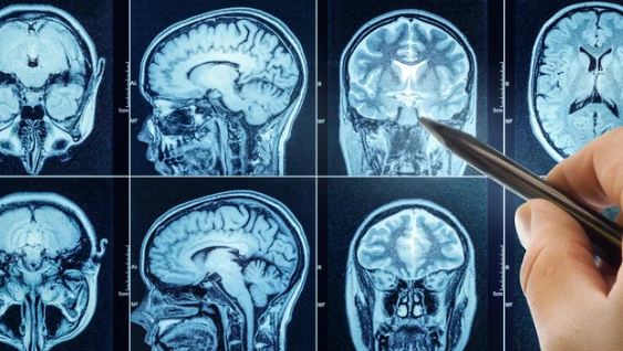
MRI scans provide detailed images of the body’s internal structures, but these images can be hard for patients to understand. Personalized visualization reports break down complex scan results into clear, easy-to-follow videos or graphics. These reports help patients see and understand their MRI scans in a way that makes medical information accessible and less confusing.
Using advanced AI technology, these personalized tools analyze MRI images quickly and deliver straightforward explanations without medical jargon. This makes it easier for patients and doctors to make informed decisions about treatment and next steps. Patients can also access their reports online, providing fast and convenient insight into their health.
Radiologists still play a key role by reviewing and verifying AI-generated reports to ensure accuracy. As technology improves, personalized visualization reports are becoming more common, giving patients better control over their medical information.
Key Takeways
- Personalized reports simplify complex MRI images for better patient understanding.
- AI technology speeds up interpretation and removes confusing medical terms.
- Access to visual reports online helps patients stay informed and involved.
Understanding MRI Scans
MRI scans provide detailed images of the inside of the body. They use magnetic fields and radio waves to create pictures of organs, tissues, and bones. Different types of MRI procedures focus on specific body parts or medical needs.
What Is an MRI
Magnetic Resonance Imaging (MRI) is a medical test that shows clear images of organs and tissues without using X-rays. It helps doctors see problems like tumors, injuries, or diseases inside the body.
An MRI scanner uses powerful magnets and radio waves to create images. These images are detailed, showing soft tissues better than other tests. The procedure is painless but may take 20 to 60 minutes depending on the scan type.
How MRI Technology Works
MRI machines create images by using strong magnetic fields to align hydrogen atoms in the body. When radio waves hit these atoms, they produce signals that the machine detects. A computer turns these signals into pictures.
The images show different shades based on tissue types and water content. This helps doctors tell normal tissues from abnormalities. The method involves no radiation, making it safer for frequent use.
Contrast dyes might be injected to highlight blood vessels or certain tissues. This makes it easier to spot diseases or injuries in complex areas like the brain or joints.
Types of MRI Procedures
There are various MRI types, each suit different medical needs:
- Brain MRI: Checks for brain tumors, strokes, or swelling.
- Spine MRI: Looks at discs, nerves, and spinal cord problems.
- Joint MRI: Evaluates injuries or arthritis in knees, shoulders, or hips.
- Cardiac MRI: Assesses heart structures and blood flow.
- Abdominal MRI: Examines organs like the liver, kidneys, and pancreas.
Some scans use contrast agents to improve detail. Others are specialized for children or sensitive conditions, adapting to patient comfort and diagnostic goals.
Personalized Visualization Reports Explained
These reports turn complex MRI data into clear, easy-to-understand formats. They focus on showing detailed images with tailored explanations. This makes it simpler for patients and doctors to see what the scan reveals and understand its medical significance.
Definition and Purpose
Personalized visualization reports use advanced imaging software to create detailed visual and sometimes video summaries of MRI scans. The goal is to help patients and doctors clearly see important findings without getting lost in technical language.
These reports often highlight areas of concern, explain abnormalities, and show normal anatomy using clear visuals. They aim to improve communication by making scan results more accessible, which helps patients understand their condition and treatment options better.
Key Features of Visualization Reports
Personalized reports often include animated videos, color-coded images, and simplified text explanations. They focus on key parts of the scan, like joints or specific organs, to show relevant details clearly.
Other features are:
- Automated image enhancement for clearer views
- Step-by-step walkthroughs of important findings
- Expert-reviewed interpretations for accuracy
- Accessibility via online platforms or apps
This approach reduces confusion and allows users to grasp information faster than reading raw MRI data alone.
Differences from Traditional MRI Results
Traditional MRI results usually come as written reports with technical terms and standard image slices. They can be hard for non-experts to understand, often requiring a doctor’s explanation.
In contrast, personalized visualization reports combine images with plain language and graphics. They focus on user-friendly presentation rather than medical jargon. This helps patients engage directly with their scan results and feel more confident discussing them with healthcare providers.
Benefits of Personalized Visualization for Patients
Personalized visualization of MRI scans offers clear advantages that improve how patients perceive and engage with their health information. It makes complex medical data easier to understand, supports patients in making choices about their care, and strengthens the connection between patients and their healthcare providers.
Enhanced Patient Understanding
Personalized visualization transforms traditional MRI scans into easy-to-navigate 3D images. These images show the exact size, shape, and location of an issue, making it easier for patients to grasp their condition.
By seeing their own anatomy from different angles, patients gain a clearer picture than with flat, 2D images. This can reduce confusion and anxiety caused by unclear or technical reports.
Clear visual information helps patients remember details and better understand explanations given by doctors. It bridges the gap between technical medical terms and patient knowledge, making health information more accessible.
Empowerment in Medical Decisions
When patients see detailed personalized images, they can make more informed choices about their treatment. Understanding their specific condition visually supports weighing options such as surgery, medication, or therapy.
Personalized scans also reveal potential risks and benefits in a clear way. This helps patients feel more confident about the decisions they make with their healthcare team.
This type of visualization supports shared decision-making, where the patient plays an active role. They can ask more specific questions and better express their preferences based on what they see.
Facilitating Communication With Healthcare Providers
Personalized MRI visualizations improve the dialogue between patients and clinicians. Visual tools help patients describe symptoms and concerns more accurately.
Doctors can show critical areas or treatment plans using the images. This reduces misunderstandings that might occur during verbal explanations.
These visuals also assist in educating patients about procedure steps or expected outcomes. Clear communication based on personalized visuals supports trust and cooperation throughout the care process.
How to Access and Read Your MRI Visualization Report
MRI visualization reports often come as digital files or online access links. They contain images, highlights, and explanations to help users see key findings clearly. Understanding how to receive the report, use the viewing platform, and interpret visuals is essential for making sense of the scan.
Receiving Your Report
MRI results are usually sent by email or made available through a secure patient portal. The report may arrive as a PDF, video file, or a link to an interactive website. Patients should check their spam folder if no message arrives within a few days after the scan.
Sometimes, MRI CDs come with built-in software that must be installed on a computer to view images. Hospitals often provide instructions for downloading apps or logging into online portals to access these reports. It is important to use a secure network when opening these files to protect private health information.
Navigating the Visualization Platform
Most MRI visualization platforms include menus for selecting different views, such as axial, coronal, and sagittal planes. Users can zoom in and out, toggle labels on or off, and sometimes view 3D reconstructions of the scanned area.
Tools like sliders or arrows allow moving through image slices one by one. Some platforms include simple explanations alongside images or customizable settings to adjust brightness and contrast. Familiarizing oneself with the controls is key to exploring images effectively.
Interpreting Common Visual Elements
MRI reports often highlight areas of interest using colors, arrows, or labels. Bright spots may indicate tissue changes; dark areas might show fluid or empty spaces. Annotations often identify normal anatomy versus anomalies.
Legends or keys explain symbols and shading used in the images. It is common to see measurements of lesions or abnormalities indicated next to the visuals. Understanding these elements helps patients grasp the size, location, and nature of any findings.
Technological Advances Behind MRI Visualization
Modern MRI visualization relies on powerful software, seamless data integration, and strong security measures. These advances help doctors and patients understand scans more clearly and keep personal information safe.
3D Imaging Software
3D imaging software transforms flat MRI images into detailed, interactive models. This allows doctors to view organs and tissues from multiple angles. It improves diagnosis by showing structures more clearly than traditional 2D images.
The software uses strong computers and smart algorithms to rebuild the body’s shape in three dimensions. It also supports zooming and rotating, giving a better sense of depth. These tools help surgeons plan operations and explain conditions to patients.
Some platforms include color coding to highlight different tissues or problems. This detailed visualization makes it easier to spot small abnormalities that might otherwise be missed.
Integration With Electronic Health Records
MRI visualization tools now connect directly with Electronic Health Records (EHR). This integration allows doctors to access scans alongside other patient data like lab results, medications, and medical history.
It streamlines workflows by reducing the need to switch between multiple systems. Doctors can review MRI reports and images within a single platform. This saves time and reduces the risk of errors in interpreting data.
This integration also supports personalized visualization reports that include tailored notes based on the patient’s unique health information. It improves communication between specialists and primary care doctors managing ongoing care.
Security and Data Privacy
Protecting patient data in MRI visualization is critical. Hospitals use encryption to secure images and reports during storage and transmission. This prevents unauthorized access or tampering.
Access controls ensure only authorized personnel can view sensitive information. Multi-factor authentication adds extra security for users logging into MRI systems or EHR platforms.
Strict privacy rules comply with healthcare regulations. These measures also include audit logs that track who accessed or modified MRI data, helping detect any suspicious activity.
By balancing easy access with strong security, providers protect patients’ privacy while delivering high-quality imaging services.
The Role of Radiologists in Personalized Reporting
Radiologists play a crucial role in making scan results clear and useful for both doctors and patients. They work closely with healthcare teams and adjust reports to fit each patient’s unique situation. This improves understanding and helps guide decisions.
Collaborative Interpretation
Radiologists do more than just read MRI scans; they interpret images alongside referring physicians to ensure accurate diagnoses. This teamwork helps clarify complex findings and connects imaging results to the patient’s overall health.
They often discuss cases with specialists to decide on the best care path. This collaboration ensures reports are precise and clinically relevant. It also supports faster, informed decisions by the medical team.
Interactive tools are increasingly used to link images to reports, making it easier for doctors to see important details. These connections improve communication and reduce the chance of misinterpretation.
Customization for Individual Patients
Radiologists tailor reports to meet each patient’s specific needs. This includes simplifying language to help patients understand their scans better, sometimes with the help of AI technologies.
Reports may highlight key findings directly relevant to a patient’s condition, avoiding unnecessary medical jargon. This makes the information accessible to non-specialists.
Customization also means adjusting the level of detail based on the patient’s medical history and the reason for the scan. Personalized visuals and summaries enhance patient engagement and support clearer conversations between doctors and patients.
Future Trends in MRI and Personal Visualization Reports
Advances in MRI technology are focused on improving accuracy, speeding up interpretation, and making scan results more useful for both doctors and patients. New tools aim to analyze images more deeply and help predict health outcomes, while also encouraging patients to understand their scans better.
Artificial Intelligence Applications
AI is becoming a major part of how MRI scans are read and analyzed. It helps doctors quickly identify abnormalities, such as tumors or lesions, with greater accuracy than before.
Machine learning models can also highlight subtle changes in scans that humans might miss. This leads to faster diagnoses and more precise treatment plans.
AI tools are integrated into MRI machines and software to automate report generation. They can summarize findings and suggest follow-up steps, saving time for radiologists.
Still, human expertise remains key. AI supports, but does not replace, doctors in interpreting images and considering patient history.
Predictive Analytics in Imaging
Predictive analytics uses data from MRI scans combined with patient details to forecast disease progression and treatment responses.
By analyzing trends and patterns within images, this approach helps identify risks early, such as the likelihood of cancer growth or neurological decline.
Hospitals and clinics use these insights to plan personalized care. For example, deciding who may benefit from surgery versus less invasive treatments.
This trend is supported by growing MRI capabilities like functional MRI (fMRI), which shows brain activity, and higher resolution imaging that improves prediction quality.
Potential for Greater Patient Engagement
New MRI reporting techniques aim to make results easier for patients to understand.
Personalized visualization reports include annotated images and clear summaries that explain what was found, using simple language and visuals.
Some systems offer interactive reports where patients can explore their scans on tablets or websites, increasing involvement in their care.
These tools can enhance communication between doctors and patients, helping patients make informed choices about their treatment.
Engaging patients through better reports has the potential to improve treatment adherence and satisfaction.
Also Read :
