Magnetic Resonance Imaging (MRI) has become one of the most powerful tools in modern medicine. Its ability to capture highly detailed images of internal organs and tissues without exposing patients to harmful radiation makes it especially valuable in diagnosing complex conditions like cancer. But how exactly does MRI work, and why is it often preferred when detecting tumors? Let’s dive deeper into this revolutionary medical imaging technology and explore its role in cancer detection.
Understanding MRI: The Basics
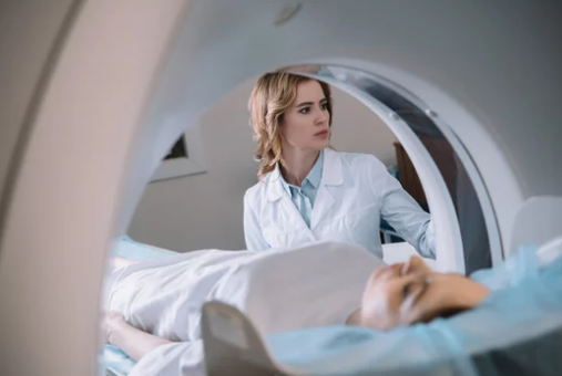
MRI, or Magnetic Resonance Imaging, is a non-invasive diagnostic technique that uses powerful magnets and radio waves to create detailed pictures of the inside of the body. Unlike X-rays or CT scans, MRI does not use ionizing radiation, making it safer for repeated use.
At its core, MRI relies on hydrogen atoms in the body. Since the human body is mostly water, and water molecules contain hydrogen, this technology is particularly effective in producing clear images of soft tissues such as the brain, muscles, heart, and internal organs.
When a patient undergoes an MRI, they lie inside a large tube-shaped machine. The scanner generates a magnetic field that temporarily aligns hydrogen atoms. Radiofrequency pulses then disrupt this alignment, and as the atoms return to their natural state, they emit signals. These signals are captured and processed by a computer to produce highly detailed cross-sectional images.
Why MRI Is Essential in Detecting Cancer
Cancer diagnosis often requires imaging that reveals both the size and location of tumors, as well as how the disease affects surrounding tissues. MRI plays a crucial role in this process for several reasons:
1. Superior Soft Tissue Contrast
MRI offers unmatched clarity when it comes to differentiating between healthy and diseased tissues. This is especially important in cancers of the brain, liver, pancreas, prostate, and soft tissues, where early detection is critical.
2. Multi-Planar Imaging
Unlike other imaging methods, MRI provides three-dimensional images from multiple angles. This allows doctors to analyze tumors in greater detail, leading to more accurate diagnoses and better treatment planning.
3. Functional Imaging Capabilities
Advanced MRI techniques, such as functional MRI (fMRI) and diffusion-weighted imaging (DWI), go beyond structural imaging. They can reveal how tissues function, how blood flows through tumors, and even identify the aggressiveness of certain cancers.
4. No Radiation Exposure
Many cancer patients undergo multiple scans during diagnosis, treatment, and follow-up. Since MRI uses no ionizing radiation, it is safer for repeated monitoring compared to CT scans or X-rays.
Types of MRI Used in Cancer Detection
Different forms of MRI are used depending on the type of cancer being investigated. Some of the most common include:
- Standard MRI – Produces detailed anatomical images of soft tissues.
- Contrast-Enhanced MRI – Uses a contrast agent, often gadolinium, to highlight abnormal tissues and improve detection accuracy.
- Functional MRI (fMRI) – Often used in brain cancer detection, fMRI maps brain activity by measuring blood flow.
- Diffusion-Weighted MRI (DW-MRI) – Helps identify cellular changes that are common in tumor development.
- Magnetic Resonance Spectroscopy (MRS) – Analyzes chemical changes in tissues, useful in distinguishing between cancerous and non-cancerous growths.
Cancers Commonly Detected with MRI
While MRI is not always the first imaging choice for every cancer, it is particularly useful for detecting:
- Brain Cancer – Provides detailed images of brain structures and helps map tumor growth.
- Breast Cancer – Often used for women at high risk or when mammograms are inconclusive.
- Prostate Cancer – Multiparametric MRI (mpMRI) has revolutionized prostate cancer detection by reducing unnecessary biopsies.
- Liver and Pancreatic Cancer – Offers precise imaging of abdominal organs, aiding in treatment planning.
- Bone and Soft Tissue Tumors – Helps distinguish between benign and malignant growths.
The Role of Contrast Agents in MRI
In many cancer-related MRIs, contrast agents are used to enhance visibility. Gadolinium-based contrast agents highlight abnormal tissue, making tumors stand out more clearly against healthy tissue. While generally safe, some patients may experience mild side effects like nausea or headaches, and those with kidney issues need careful evaluation before use.
MRI vs. Other Imaging Techniques in Cancer Detection
To understand MRI’s advantages, it’s helpful to compare it with other common imaging methods:
- X-rays – Best for bone imaging but not effective for soft tissue cancers.
- CT Scans – Faster and widely available but involve radiation exposure.
- PET Scans – Excellent for showing cancer activity but usually paired with CT or MRI for anatomical accuracy.
- Ultrasound – Useful for certain cancers like breast and liver but lacks MRI’s detailed resolution.
Overall, MRI stands out for its ability to capture precise, radiation-free images, making it an indispensable tool in cancer diagnosis.
Limitations of MRI in Cancer Detection
Despite its advantages, MRI is not perfect. Some limitations include:
- High Cost – MRI scans are more expensive than CT or ultrasound.
- Longer Scanning Time – Patients must remain still for 30–60 minutes, which can be uncomfortable.
- Claustrophobia Concerns – The enclosed scanner can trigger anxiety in some patients.
- Limited Availability – Not all hospitals have advanced MRI machines or specialists trained in cancer imaging.
How MRI Helps Guide Cancer Treatment
Beyond detection, MRI plays a major role in cancer treatment and follow-up care. Doctors use MRI to:
- Assess tumor response to chemotherapy or radiation.
- Plan surgeries with precision by mapping tumor boundaries.
- Monitor for cancer recurrence after treatment.
- Evaluate metastasis (cancer spread) in surrounding tissues.
This makes MRI not just a diagnostic tool but also a cornerstone in ongoing cancer management.
Future of MRI in Cancer Diagnosis
Advancements in MRI technology are rapidly transforming cancer care. Artificial intelligence (AI) integration, faster scanning techniques, and ultra-high-field MRI machines are making imaging more accurate and accessible. Researchers are also exploring how MRI biomarkers can predict cancer progression and treatment response, potentially allowing for more personalized medicine.
Conclusion
MRI is more than just a diagnostic tool—it is a lifesaving technology that has revolutionized how doctors detect, monitor, and treat cancer. Its ability to deliver detailed images of soft tissues without radiation exposure makes it one of the safest and most effective imaging techniques available today.
As technology continues to evolve, MRI will play an even greater role in early cancer detection and precision medicine. For patients and doctors alike, it represents a critical ally in the ongoing fight against cancer.
Also Read :