Cancer is one of the most challenging diseases to diagnose and treat, and early detection often makes a life-saving difference. Medical imaging plays a central role in identifying suspicious growths, and one of the most powerful tools available today is Magnetic Resonance Imaging (MRI). If you’re new to understanding how medical imaging helps in cancer care, this beginner’s guide will walk you through how MRI works, why it’s used, and what to expect during the process.
What Is MRI?
MRI, short for Magnetic Resonance Imaging, is a non-invasive medical imaging technique that uses strong magnets, radio waves, and computer technology to create detailed pictures of the inside of the body. Unlike X-rays or CT scans, MRI does not use harmful ionizing radiation, which makes it a safer option for repeated scans.
The key advantage of MRI is its ability to show soft tissues—such as the brain, muscles, heart, and internal organs—with incredible clarity. This makes it especially valuable in detecting cancers that may not be visible on other types of scans.
How Does MRI Work?
At first, MRI might seem like complex science, but the principle is straightforward when broken down:
- Magnetic Field Alignment
The MRI machine contains a very strong magnet. When you lie inside the scanner, the magnetic field temporarily aligns hydrogen atoms in your body. Since the human body is made mostly of water, and water contains hydrogen, this alignment is possible. - Radiofrequency Pulses
The scanner then sends out radiofrequency (RF) pulses, which momentarily knock these hydrogen atoms out of alignment. - Signal Emission
As the hydrogen atoms return to their natural state, they release tiny signals. - Image Creation
The MRI computer captures these signals and processes them into highly detailed cross-sectional images of tissues and organs.
The result? Doctors get an in-depth view of your internal body structures without any cutting, surgery, or radiation exposure.
Why MRI Is Important in Cancer Diagnosis
Cancer diagnosis isn’t just about spotting a tumor—it’s also about understanding its size, exact location, and impact on surrounding tissues. MRI provides valuable insights in several ways:
- High-Resolution Imaging – Detects tumors that might be missed by other imaging techniques.
- Soft Tissue Contrast – Distinguishes between healthy tissue and cancerous growths.
- Multi-Plane Views – Produces images from different angles for better analysis.
- No Radiation – Safer for patients who need multiple scans over time.
- Functional Imaging – Advanced MRI methods can even show how a tumor is behaving.
Types of MRI Used in Cancer Detection
Not all MRIs are the same. Depending on the situation, doctors may choose different MRI techniques:
- Standard MRI – Provides clear images of internal structures.
- Contrast-Enhanced MRI – Involves injecting a contrast dye (usually gadolinium) to highlight abnormal tissues.
- Functional MRI (fMRI) – Often used for brain cancers to track activity and blood flow.
- Diffusion-Weighted Imaging (DWI) – Detects changes at the cellular level, useful in spotting tumors.
- Magnetic Resonance Spectroscopy (MRS) – Analyzes the chemical composition of tissues, helping to distinguish cancer from non-cancer growths.
Which Cancers Are Best Detected by MRI?
MRI is particularly effective for detecting and monitoring cancers in areas where soft tissue detail is crucial. These include:
- Brain tumors – Detailed images help in diagnosis and surgical planning.
- Breast cancer – Especially useful for women with dense breast tissue.
- Prostate cancer – Multiparametric MRI helps detect tumors and reduce unnecessary biopsies.
- Liver and pancreatic cancer – Provides clear imaging of complex abdominal organs.
- Spinal cord and bone marrow cancers – Identifies abnormalities that other scans may miss.
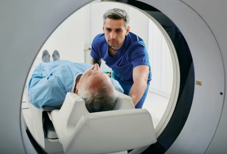
What to Expect During an MRI for Cancer Diagnosis
If you’re scheduled for an MRI, knowing what happens can help ease anxiety:
- Preparation – You may be asked to avoid eating or drinking before the scan. Remove all metal objects (jewelry, watches, hearing aids).
- Positioning – You’ll lie on a movable table that slides into the MRI machine, which looks like a large tube.
- The Scan – The machine makes loud tapping or thumping noises while it takes images. Earplugs or headphones are usually provided.
- Duration – Scans typically last 30–60 minutes, depending on the area being examined.
- Contrast Dye (if needed) – Some scans require a dye injection to highlight certain tissues. Most people tolerate this well.
- After the Scan – You can usually go home right after unless sedation was used.
Advantages of MRI in Cancer Detection
- Non-invasive and safe
- No radiation exposure
- Excellent for soft tissue imaging
- Helps guide treatment planning
- Effective for monitoring cancer progression or recurrence
Limitations of MRI
While MRI is powerful, it’s not perfect. Some challenges include:
- Higher cost compared to CT or ultrasound.
- Longer scan times, which require patients to stay very still.
- Claustrophobia – Some patients feel uncomfortable inside the narrow tube.
- Not suitable for everyone – Patients with certain implants (pacemakers, cochlear implants, or some metal fragments) may not be eligible.
- Less effective for lung or bone cancers compared to CT scans.
The Role of MRI in Cancer Treatment Planning
MRI doesn’t just detect cancer—it also guides doctors in choosing the best treatment approach. It can:
- Help surgeons map tumor boundaries before an operation.
- Track how tumors respond to chemotherapy or radiation.
- Detect whether cancer has spread to nearby tissues.
- Monitor for recurrence after treatment.
This makes MRI a key tool throughout the entire cancer journey, from diagnosis to follow-up.
Future of MRI in Cancer Diagnosis
The future looks even brighter for MRI technology. Researchers are developing faster machines, AI-powered image analysis, and hybrid imaging systems that combine MRI with PET scans for even greater accuracy. These advancements may one day make cancer detection quicker, more precise, and more accessible.
Conclusion
MRI is a powerful ally in the fight against cancer. By providing detailed, radiation-free images of the body’s soft tissues, it helps doctors detect tumors early, plan treatments effectively, and monitor recovery.
For beginners trying to understand medical imaging, think of MRI as a safe, high-tech camera that gives doctors an inside view of your body—helping ensure that cancer is caught and treated as soon as possible.
Also Read :