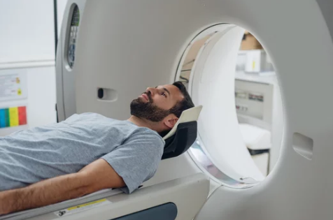
Early and accurate cancer detection is vital for effective treatment and improved patient outcomes. Among the advanced imaging techniques revolutionizing oncology, Diffusion-Weighted Imaging (DWI) has emerged as a powerful tool. DWI, a specialized MRI method, provides insights not just into the structure of tissues but also into their cellular environment and function.
This article explores how DWI works, its applications in cancer diagnosis, advantages, limitations, and why it is becoming an indispensable part of modern oncology.
What Is Diffusion-Weighted Imaging (DWI)?
Diffusion-Weighted Imaging is an MRI technique that measures the movement of water molecules within tissues. In healthy tissues, water molecules move relatively freely, but in areas with dense cellular structures, such as tumors, their movement is restricted.
This restriction generates high-contrast images that allow radiologists to identify abnormal tissue, often before it becomes visible on conventional MRI or CT scans.
How DWI Works
DWI works by detecting the Brownian motion of water molecules:
- Normal Tissue: Water diffuses freely in open, healthy tissue, resulting in low signal intensity on DWI.
- Tumor Tissue: Cancerous tissues are densely packed with cells, restricting water movement, which appears as bright areas on DWI scans.
- Quantitative Analysis: The Apparent Diffusion Coefficient (ADC) is calculated from DWI data, providing a numeric measure of water mobility. Lower ADC values often correlate with higher cellularity and malignancy.
This combination of qualitative and quantitative information makes DWI highly effective for detecting and characterizing tumors.
Applications of DWI in Cancer Diagnosis
1. Brain Tumors
- DWI is particularly useful for distinguishing high-grade from low-grade gliomas.
- Detects tumor recurrence and differentiates it from post-treatment changes, such as radiation necrosis.
2. Prostate Cancer
- DWI is a key component of multiparametric MRI (mpMRI) for prostate cancer detection.
- Helps identify clinically significant tumors, guiding targeted biopsies and reducing unnecessary procedures.
3. Liver Cancer
- Detects small hepatocellular carcinoma lesions that may be missed on standard MRI or CT.
- Monitors treatment response after interventions like ablation or chemotherapy.
4. Breast Cancer
- DWI can distinguish between benign and malignant breast lesions.
- Useful for high-risk patients and those with dense breast tissue where mammography may be less sensitive.
5. Whole-Body Oncology Imaging
- DWI is increasingly used in whole-body MRI scans to detect metastases and assess tumor burden without radiation exposure.
Advantages of DWI in Cancer Diagnosis
- Non-invasive and safe: No radiation, making it suitable for repeated imaging.
- Early detection: Can identify tumors before structural changes are visible.
- Functional information: Provides insights into tumor cellularity and aggressiveness.
- Treatment monitoring: Detects early response to chemotherapy, radiotherapy, or targeted therapies.
- Complementary to other imaging: Enhances the diagnostic power of conventional MRI or CT scans.
Limitations of DWI
While DWI is highly effective, it has some limitations:
- Sensitivity to motion: Patient movement can blur images, especially in abdominal scans.
- Artifacts: Susceptible to distortion near air-filled regions like lungs or sinuses.
- Interpretation requires expertise: ADC values can vary between scanners and protocols.
- Not definitive: DWI indicates abnormal tissue but cannot always differentiate between cancer and other causes of high cellularity, such as inflammation or infection.
DWI vs. Other Imaging Techniques
| Feature | DWI | CT Scan | Standard MRI | PET Scan |
|---|---|---|---|---|
| Radiation | None | Yes | None | Yes |
| Soft Tissue Contrast | Excellent | Moderate | Good | Moderate |
| Functional Data | Yes (water diffusion) | No | No | Yes (metabolic activity) |
| Early Detection | High | Moderate | Moderate | High |
| Whole-Body Capability | Yes (WB-MRI) | Limited | No | Yes |
DWI is particularly valuable when used alongside multiparametric MRI or PET imaging, offering both structural and functional insights.
Future Directions of DWI in Oncology
- Artificial Intelligence (AI): AI-assisted image analysis improves lesion detection and ADC interpretation.
- Faster imaging sequences: Reduces scan time while maintaining high-quality images.
- Integration with hybrid imaging: Combining DWI with PET/MRI provides both metabolic and cellular information.
- Predictive biomarkers: ADC values may help predict tumor aggressiveness and treatment response before clinical changes occur.
Conclusion
Diffusion-Weighted Imaging is revolutionizing cancer diagnosis by providing functional insights into tumor biology that conventional imaging cannot offer. From early detection to monitoring therapy and identifying metastases, DWI enhances the accuracy and effectiveness of modern oncology.
While it is not a standalone diagnostic tool, when combined with other imaging techniques, DWI provides a comprehensive and non-invasive approach to cancer care, helping clinicians detect tumors earlier, plan treatments more precisely, and monitor patient progress more effectively.
Also Read :
