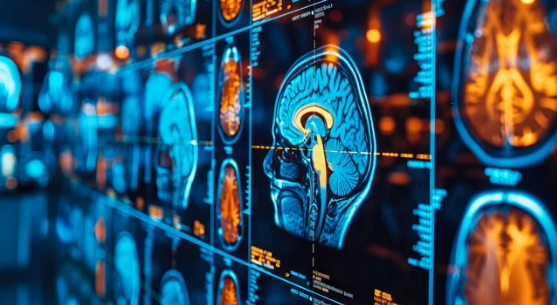
Radiation therapy is one of the most common and effective treatments for cancer. Its goal is to deliver powerful beams of radiation that destroy cancer cells while sparing as much healthy tissue as possible. Achieving this delicate balance requires precise planning—and that’s where Magnetic Resonance Imaging (MRI) plays a vital role.
MRI has become an essential tool in radiation therapy planning because of its ability to provide highly detailed, three-dimensional views of tumors and surrounding tissues. By integrating MRI into the planning process, doctors can design more accurate and personalized treatment plans for each patient.
Why Imaging Matters in Radiation Therapy
Radiation therapy relies on pinpoint accuracy. The radiation beam must:
- Target the tumor exactly
- Avoid damaging nearby healthy organs and tissues
- Deliver the correct dose for maximum effectiveness
If the tumor’s location, size, or shape is not mapped correctly, the treatment may be less effective or cause unnecessary side effects. Traditional imaging tools like CT scans provide useful information, but MRI offers a sharper and more detailed picture—especially of soft tissues such as the brain, prostate, and pelvis.
How MRI Improves Radiation Therapy Planning
1. Defining Tumor Boundaries
MRI provides clearer images of soft tissues than CT scans. This makes it easier for doctors to see the exact borders of a tumor. For example:
- In brain cancer, MRI helps distinguish tumor tissue from healthy brain tissue.
- In prostate cancer, MRI shows precise tumor location, helping doctors protect nearby organs like the bladder and rectum.
2. Mapping Organs at Risk (OARs)
Radiation must be carefully shaped to avoid harming critical organs near the tumor. MRI identifies these structures with great accuracy, allowing doctors to design treatment beams that reduce side effects.
3. Functional Imaging for Better Targeting
Advanced MRI techniques provide information beyond anatomy:
- Diffusion-Weighted Imaging (DWI): Shows how water molecules move in tissues, helping detect cancer aggressiveness.
- Functional MRI (fMRI): Maps areas of the brain responsible for speech, vision, and movement to avoid damage during radiation therapy.
- Dynamic Contrast-Enhanced MRI (DCE-MRI): Measures blood flow, showing how tumors interact with surrounding tissues.
4. Combining MRI with CT for Planning
In many cancer centers, CT scans are still used to calculate radiation doses. However, when MRI images are combined with CT scans, doctors get the best of both worlds:
- CT for dose calculation
- MRI for precise tumor and organ visualization
This fusion creates a more accurate treatment plan.
MRI-Guided Radiation Therapy: Real-Time Imaging
A groundbreaking advancement is MRI-guided radiation therapy. This technology allows doctors to see the tumor in real time while delivering radiation.
How It Works
- The patient lies on a treatment table inside an MRI machine equipped with a radiation system.
- MRI continuously monitors the tumor’s position and nearby organs.
- If the tumor moves (for example, due to breathing), the system adjusts the radiation beam instantly.
Benefits of MRI-Guided Therapy
- Improved accuracy in targeting moving tumors (like lung or liver cancers).
- Adaptive therapy: Doctors can adjust the treatment plan day by day based on new MRI images.
- Reduced side effects by sparing more healthy tissue.
Types of Cancers That Benefit Most from MRI in Radiation Therapy
- Brain tumors: MRI identifies tumor boundaries and critical brain functions.
- Prostate cancer: MRI shows tumor location and guides precise targeting.
- Breast cancer: Helps define tumor margins and avoid damage to the heart and lungs.
- Cervical and other pelvic cancers: MRI maps tumor spread and nearby reproductive organs.
- Head and neck cancers: MRI improves visualization of complex structures.
Advantages of Using MRI for Radiation Planning
- More accurate tumor visualization
- Better protection for nearby healthy tissues
- Ability to adapt treatment based on daily changes
- Enhanced detection of microscopic tumor spread
- Reduced risk of long-term complications
Limitations and Challenges
While MRI is powerful, it does have some challenges:
- Availability: Not all cancer centers have MRI-guided radiation systems.
- Cost: MRI-guided therapy can be more expensive.
- Time: MRI scans may take longer than CT scans.
- Technical expertise: Requires specialized teams for planning and execution.
Future of MRI in Radiation Therapy
The integration of MRI into cancer treatment is rapidly evolving. Future advancements may include:
- AI-assisted MRI planning: Using artificial intelligence to automate and improve accuracy.
- Faster MRI scans: To shorten treatment times.
- Personalized adaptive therapy: Daily MRI-based adjustments tailored to each patient’s anatomy and tumor response.
- MRI combined with proton therapy: Offering even greater precision and reduced side effects.
Final Thoughts
MRI is transforming how doctors plan and deliver radiation therapy. By providing highly detailed images of tumors and surrounding tissues, it ensures that radiation beams hit their target with unmatched precision. For patients, this means more effective treatment, fewer side effects, and improved quality of life.
As MRI-guided radiation therapy becomes more widely available, it promises to make cancer treatment more personalized and adaptive—helping patients receive the right dose, at the right time, in exactly the right place.
Also Read :
