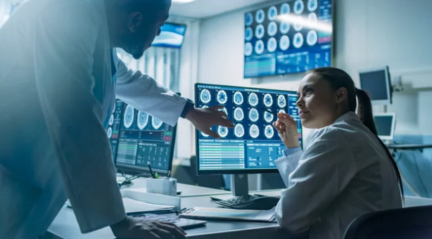
Completing cancer treatment is a significant milestone, but the journey doesn’t end there. Even after surgery, chemotherapy, or radiation therapy, patients need ongoing monitoring to ensure the cancer hasn’t returned and that no hidden complications are developing. One of the most valuable tools in this stage of care is Magnetic Resonance Imaging (MRI).
MRI is often used in post-treatment monitoring because of its ability to provide clear, detailed images of the body without radiation exposure. This makes it especially suitable for long-term follow-up, where multiple scans may be needed over months or years.
Why Post-Treatment Monitoring Matters
After treatment, doctors must carefully track a patient’s health to:
- Detect cancer recurrence early
- Monitor for metastasis (spread of cancer)
- Check if any residual tumor tissue remains
- Assess treatment side effects or complications
- Support long-term recovery and quality of life
Early detection during follow-up can make a huge difference. If a recurrence is found at an early stage, patients often have more treatment options and better outcomes.
Why MRI Is Preferred for Post-Treatment Monitoring
MRI stands out compared to other imaging methods like CT scans or X-rays because of several advantages:
- No radiation exposure: Unlike CT scans, MRI uses magnetic fields and radio waves, making it safe for repeated scans.
- Excellent soft tissue contrast: MRI provides highly detailed images of organs, muscles, the brain, and other soft tissues.
- Advanced functional imaging: Techniques like diffusion-weighted imaging (DWI) and dynamic contrast-enhanced MRI (DCE-MRI) reveal how tissues are behaving, not just how they look.
- Whole-body imaging: For patients at risk of metastasis, whole-body MRI can scan multiple areas at once.
How MRI Is Used in Post-Treatment Monitoring
1. Detecting Recurrence
MRI is highly sensitive in spotting even small lesions that may indicate cancer has returned. For example:
- In breast cancer, MRI can detect new tumors not visible on mammography.
- In brain tumors, MRI reveals recurrence with more accuracy than CT scans.
2. Checking for Residual Disease
Sometimes treatment doesn’t completely remove or destroy all cancer cells. MRI helps identify whether any tumor tissue remains, guiding further therapy if needed.
3. Monitoring Treatment Effects
Cancer treatment itself can cause changes in organs and tissues. MRI helps monitor:
- Scarring or fibrosis after radiation therapy
- Changes in blood supply to organs
- Side effects like swelling or nerve damage
4. Functional and Metabolic Insights
With advanced imaging techniques, MRI can track biological activity:
- DWI detects changes in how water molecules move in tissues, which may indicate early recurrence.
- MRS (Magnetic Resonance Spectroscopy) analyzes chemical changes within tissues, useful in brain and prostate cancer follow-up.
5. Whole-Body MRI for Metastasis
Whole-body MRI is increasingly used to check whether cancer has spread to bones, liver, lungs, or other organs, all in one scan session.
Types of Cancer Where MRI Is Commonly Used for Monitoring
- Brain tumors: MRI is the gold standard for long-term follow-up.
- Breast cancer: Especially for high-risk patients or those with dense breast tissue.
- Prostate cancer: Multiparametric MRI provides detailed evaluation of possible recurrence.
- Liver cancer: MRI detects recurrence and assesses post-treatment liver function.
- Bone and soft tissue cancers: Whole-body MRI helps track metastasis.
Benefits for Patients
- Safety: No cumulative radiation risk, making it safe for repeated use.
- Accuracy: Detects small or hidden recurrences early.
- Peace of mind: Provides reassurance during survivorship care.
- Personalized care: Helps doctors tailor follow-up and additional treatments if needed.
Limitations of MRI in Post-Treatment Care
- Availability and cost: MRI machines and specialized imaging techniques may not be accessible everywhere.
- Contrast dye concerns: Some scans require gadolinium-based contrast, which may not be suitable for patients with kidney problems.
- Longer scan times: MRI can take 30–60 minutes, which may be uncomfortable for some patients.
- Interpretation complexity: Requires highly skilled radiologists to distinguish between scar tissue and true recurrence.
The Future of MRI in Post-Treatment Monitoring
Advances in MRI technology are making follow-up care even more effective:
- AI-assisted analysis: Artificial intelligence is helping radiologists detect subtle changes faster and with greater accuracy.
- Faster whole-body MRI scans: New techniques are reducing scan time while keeping detail.
- Molecular and functional imaging: Offering insights into cancer biology, not just structure.
- Integration with blood biomarkers: MRI combined with liquid biopsies may provide a more complete picture of recurrence risk.
Final Thoughts
MRI has become one of the most trusted tools for post-treatment cancer monitoring. By offering safe, detailed, and functional imaging, it allows doctors to detect recurrence early, monitor treatment effects, and ensure the best possible long-term care for patients.
For cancer survivors, MRI is more than just a scan—it is an important part of the journey toward recovery, reassurance, and continued health.
Also Read :