When it comes to diagnosing cancer, accuracy is everything. A biopsy—where doctors take a small sample of tissue for examination under a microscope—is often the only way to confirm whether a suspicious lump or lesion is cancerous. But not all biopsies are the same. Traditionally, biopsies are guided using imaging techniques like ultrasound or CT scans. However, MRI-guided biopsies are gaining attention for their higher precision, especially in complex cases.
In this article, we’ll explore how MRI-guided biopsies work, their advantages over other methods, and whether they truly provide more accuracy for cancer diagnosis.
What Is an MRI-Guided Biopsy?
An MRI-guided biopsy is a procedure where doctors use Magnetic Resonance Imaging (MRI) to guide the removal of tissue samples from a suspicious area.
Unlike ultrasound or CT scans, MRI offers:
- Superior soft tissue detail
- Multiplanar imaging (3D perspectives)
- Functional insights like blood flow and tissue density
This makes MRI particularly valuable when dealing with tumors that are hard to detect or poorly defined with other imaging tools.
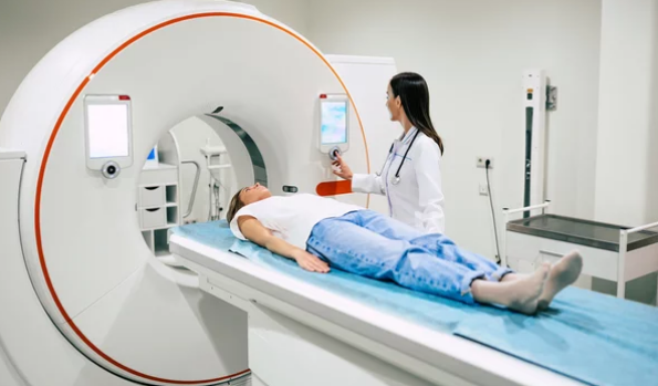
How the Procedure Works
- Patient Preparation
The patient lies inside an MRI scanner, usually in a position that gives the clearest view of the suspicious tissue. - Initial MRI Scan
Doctors perform an initial scan to precisely locate the lesion. - Targeting the Area
Using MRI images, the radiologist identifies the exact location and angle for the biopsy needle. - Needle Insertion
A special MRI-compatible needle is inserted into the targeted tissue under continuous imaging guidance. - Sample Collection
Tissue samples are collected and sent to a pathology lab for microscopic examination. - Post-Procedure Scan
A final MRI confirms that the correct tissue was sampled and checks for complications such as bleeding.
Why MRI-Guided Biopsies Are More Accurate
1. Superior Imaging for Hard-to-See Tumors
MRI is particularly effective for areas where ultrasound and CT scans struggle, such as:
- Prostate gland (deep tissue, difficult to visualize with ultrasound alone)
- Breast tissue (especially dense breast tissue)
- Brain and spinal cord lesions
- Liver tumors hidden by surrounding tissue
2. Better Tumor Characterization
Advanced MRI techniques like Diffusion-Weighted Imaging (DWI) and Dynamic Contrast-Enhanced MRI (DCE-MRI) help differentiate between suspicious and benign tissues. This allows radiologists to target the most concerning areas for biopsy.
3. Reduced Sampling Errors
Conventional biopsies may miss the tumor, especially if it’s small or irregularly shaped. MRI guidance ensures the needle reaches the exact spot, reducing the chance of false negatives.
4. Useful in Previous Negative Biopsies
In cases where ultrasound- or CT-guided biopsies fail to find cancer despite strong clinical suspicion, MRI-guided biopsies often succeed.
Examples of Cancers Where MRI-Guided Biopsies Are Used
- Prostate Cancer: Multiparametric MRI (mpMRI) combined with targeted biopsy increases detection rates of clinically significant cancers.
- Breast Cancer: MRI-guided biopsy is vital when lesions are visible on MRI but not on mammography or ultrasound.
- Brain Tumors: MRI guides surgeons to sample precise tumor regions while avoiding critical brain functions.
- Liver Cancer: MRI identifies small or hidden lesions for targeted biopsy.
Benefits for Patients
- Higher diagnostic accuracy
- Fewer repeat biopsies needed
- Greater confidence in treatment planning
- Early detection of aggressive cancers that may otherwise be missed
Limitations and Challenges
Despite its benefits, MRI-guided biopsies also come with drawbacks:
- Time-consuming: The procedure takes longer than ultrasound-guided biopsies.
- Cost: MRI machines and specialized equipment make it more expensive.
- Limited availability: Not all hospitals have MRI-guided biopsy capabilities.
- Patient comfort: Lying still inside an MRI scanner for a long time may be uncomfortable.
Future of MRI-Guided Biopsies
Ongoing research is making these biopsies even more effective:
- Fusion imaging: Combining MRI with ultrasound for faster and more cost-effective biopsies.
- AI-assisted targeting: Artificial intelligence helping radiologists pinpoint suspicious regions more accurately.
- Molecular MRI: Detecting tumors at the cellular level for earlier diagnosis.
Final Thoughts
So, are MRI-guided biopsies more accurate? Yes—especially in cases where traditional methods fall short. They provide unmatched precision, reduce false negatives, and improve detection of cancers that might otherwise remain hidden.
While not always necessary for every patient due to cost and availability, MRI-guided biopsies are a powerful option when accuracy is critical. For cancers like prostate, breast, and brain tumors, they are increasingly becoming the gold standard in diagnosis.
By improving biopsy accuracy, MRI guidance not only enhances cancer detection but also ensures patients receive the right treatment sooner—offering hope for better outcomes and survival rates.
Also Read :