Cancer surgery has always required a delicate balance: removing as much tumor tissue as possible while preserving healthy organs and tissues. Traditionally, surgeons have relied on preoperative imaging and their experience to guide procedures. However, real-time MRI during cancer surgery is revolutionizing this process, offering unprecedented precision and transforming surgical outcomes.
This article explores the concept of intraoperative MRI, its benefits, applications in various cancers, and why it represents a major advancement in personalized cancer care.
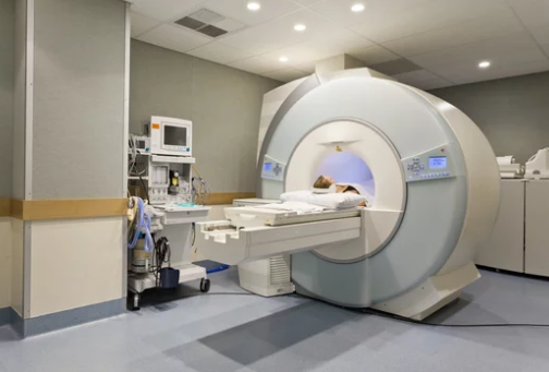
What Is Real-Time MRI During Surgery?
Real-time, or intraoperative, MRI (iMRI) is an imaging technique that allows surgeons to view high-resolution MRI scans while the patient is still in the operating room. Unlike traditional MRI performed before surgery, iMRI provides continuous imaging updates, enabling the surgical team to adjust procedures on the fly.
This capability is particularly important in complex surgeries where tumor boundaries are unclear or located near critical structures such as the brain, spinal cord, or vital organs.
How Real-Time MRI Works
- Preoperative Planning
Before surgery, standard MRI scans map the tumor’s location, size, and surrounding anatomy. Surgeons use these images to develop an initial surgical plan. - Intraoperative Imaging
During the procedure, the patient is positioned in an MRI-compatible operating room or scanner. MRI images are acquired in real time, allowing the surgeon to see tumor tissue, residual cancer, and critical structures. - Dynamic Adjustments
If the surgeon discovers remaining tumor tissue or unexpected anatomy, the iMRI provides immediate feedback. This enables precise removal while minimizing damage to healthy tissue. - Post-Resection Verification
A final intraoperative MRI scan confirms complete tumor removal and ensures surgical margins are clear, reducing the likelihood of repeat surgeries.
Advantages of Real-Time MRI in Cancer Surgery
1. Increased Surgical Precision
iMRI allows surgeons to differentiate between tumor tissue and healthy tissue with unparalleled clarity. This is especially important for tumors in the:
- Brain: Avoiding critical regions responsible for speech, movement, or vision
- Spine: Preventing damage to the spinal cord
- Liver: Protecting major blood vessels during resection
2. Reduced Need for Reoperations
Residual tumor tissue after surgery is a major cause of repeat procedures. iMRI helps confirm complete removal during the initial surgery, lowering the risk of recurrence.
3. Better Preservation of Healthy Tissue
By visualizing critical structures in real time, surgeons can avoid unnecessary damage, improving post-surgical function and quality of life.
4. Personalized Surgical Approach
iMRI supports tailored surgical plans based on each patient’s anatomy and tumor characteristics, contributing to the broader goal of personalized cancer care.
Applications of Real-Time MRI in Cancer Surgery
1. Brain Tumors
iMRI is widely used for gliomas and other brain tumors, where distinguishing tumor tissue from healthy brain is challenging. It helps minimize neurological damage while achieving maximal tumor removal.
2. Prostate Cancer
For prostatectomy procedures, iMRI guides surgeons to remove cancerous tissue precisely, sparing nerves responsible for urinary and sexual function.
3. Liver and Pancreatic Tumors
iMRI assists in identifying tumor margins near major blood vessels, reducing complications and ensuring complete resection.
4. Spine Tumors
Surgeons can safely remove spinal tumors while avoiding damage to nerves and vertebral structures, minimizing long-term disability.
Benefits for Patients
- Higher success rates: More complete tumor removal with fewer complications
- Fewer repeat surgeries: iMRI confirms tumor resection in real time
- Preserved organ function: Minimizing damage to healthy tissue improves quality of life
- Personalized care: Each surgery is tailored to the patient’s unique anatomy and tumor characteristics
Challenges and Considerations
While promising, iMRI also presents challenges:
- Cost: MRI-compatible operating rooms and equipment are expensive.
- Technical complexity: Requires specialized surgical teams trained in MRI-guided procedures.
- Longer surgery times: Scanning and positioning may increase operative duration.
- Availability: Not all hospitals have iMRI capabilities, limiting access.
The Future of Real-Time MRI in Cancer Surgery
The integration of iMRI with emerging technologies promises even greater advances:
- AI-assisted imaging: Artificial intelligence can highlight tumor margins and critical structures in real time.
- MRI-guided robotic surgery: Combining real-time imaging with robotic precision for minimally invasive procedures.
- Advanced functional MRI: Mapping organ function during surgery for safer tumor removal.
- Global accessibility: Portable and lower-cost MRI solutions may expand availability worldwide.
Conclusion
Real-time MRI during cancer surgery represents a revolutionary step in precision medicine. By providing high-resolution, live imaging in the operating room, it allows surgeons to remove tumors more completely, protect healthy tissue, and tailor procedures to each patient’s unique anatomy.
As technology continues to advance with AI integration, robotic surgery, and functional imaging, iMRI is poised to become a standard in complex cancer surgeries—offering patients safer procedures, better outcomes, and a higher quality of life.
Also Read :